Patent application title: METHOD FOR THE INHIBITION OF ANGIOGENESIS
Inventors:
Marsha A. Moses (Brookline, MA, US)
Matthew J. Foradori (Edinboro, PA, US)
Robert S. Langer (Newton, MA, US)
Assignees:
CHILDREN'S MEDICAL CENTER CORPORATION
IPC8 Class: AA61K3817FI
USPC Class:
514 12
Class name: Designated organic active ingredient containing (doai) peptide containing (e.g., protein, peptones, fibrinogen, etc.) doai 25 or more peptide repeating units in known peptide chain structure
Publication date: 2009-08-13
Patent application number: 20090203590
Inventors list |
Agents list |
Assignees list |
List by place |
Classification tree browser |
Top 100 Inventors |
Top 100 Agents |
Top 100 Assignees |
Usenet FAQ Index |
Documents |
Other FAQs |
Patent application title: METHOD FOR THE INHIBITION OF ANGIOGENESIS
Inventors:
Robert S. Langer
Marsha A. Moses
Matthew J. Foradori
Agents:
DAVID S. RESNICK
Assignees:
CHILDREN'S MEDICAL CENTER CORPORATION
Origin: BOSTON, MA US
IPC8 Class: AA61K3817FI
USPC Class:
514 12
Abstract:
The present invention is based on the discovery that Matrilin-1 has
antiangiogenic and anticancer properties. The invention is directed to a
method of treating a disease that responds to an inhibition of
angiogenesis. Additionally, the invention can be applied to those at risk
for developing a disease that responds to an inhibition of angiogenesis.
The methods comprise administering to a mammal an effective
angiogenesis-inhibiting amount of an Matrilin-1 consisting of Matrilin-1,
Matrilin-1 fragment, analog, or derivative that is administered in a
composition substantially free of other cartilage proteins.Claims:
1. A method for treatment of a disease that responds to all inhibition of
angiogenesis comprising administering to a mammal a pharmaceutical
composition comprising an effective angiogenesis-inhibiting amount of a
Matrilin-1, wherein the composition is substantially free of other
cartilage proteins.
2. The method of claim 1, wherein the disease that responds to an inhibition of angiogenesis is cancer.
3. The method of claim 1, wherein Matrilin-1 is human Matrilin-1 and comprises amino acids 1-496 of SEQ ID NO.: 1.
4. The method of claim 3, wherein Matrilin-1 comprises amino acids 23-222 of SEQ ID NO.: 1.
5. The method of claim 3, wherein Matrilin-1 comprises amino acids 43-222 of SEQ ID NO.: 1.
6. The method of claim 3, wherein Matrilin-1 comprises amino acids 264-453 of SEQ ID NO.: 1.
7. The method of claim 3, wherein Matrilin-1 comprises amino acids 278-453 SEQ ID NO.: 1.
8. The method of claim 1, wherein Matrilin-1 is Shark Matrilin-1.
9. The method of claim 1, wherein Matrilin-1 is Chick Matrilin-1 and comprises amino acids 1-493 of SEQ ID NO.: 2.
10. The method of claim 9, wherein Matrilin-1 comprises amino acids 24-220 of SEQ ID NO.: 2.
11. The method of claim 9, wherein Matrilin-1 comprises amino acids 42-220 of SEQ ID NO.: 2.
12. The method of claim 9, wherein Matrilin-1 comprises amino acids 262-450 of SEQ ID NO.: 2.
13. The method of claim 9, wherein Matrilin-1 comprises amino acids 275-450 of SEQ ID NO.: 2.
14. The method of claim 1, wherein Matrilin-1 comprises a Matrilin-1 derivative having at least 50% identity compared to a fragment of Matrilin-1 from which the derivative was derived, wherein the derivative is derived from SEQ ID NO: 1, or SEQ ID NO: 2.
15. The method of claim 1, wherein Matrilin-1 is recombinant Matrilin-1.
16. The method of claim 1, wherein said disease that responds to an inhibition of angiogenesis is retinopathy of prematurity, diabetic retinopathy or macular degeneration.
17. The method of claim 1, wherein said disease the responds to an inhibition of angiogenesis is arthritis or rheumatoid arthritis.
18. The method of claim 1, wherein said administering is conducted in conjunction with chemotherapy.
19. The method of claim 1, wherein said administering is conducted in conjunction with radiation therapy.
20. The method of claim 1, wherein said administering is conducted in conjunction with a second angiogenesis inhibitor.
21. The method of claim 1, wherein said mammal is at risk for developing said disease the responds to an inhibition of angiogenesis.
22. The method of claim 1, wherein said Matrilin-1 is incorporated into a stent for local release and inhibition of restenosis.
23. A medical device comprising Matrilin-1.
24. The medical device of claim 1, wherein the device is a stent, catheter, cannula, or an electrode.
25. A pharmaceutical composition comprising Matrilin-1, wherein the composition is substantially free of other cartilage proteins.
26. The composition of claim 25, wherein the composition is formulated for sustained release.
27. The composition of claim 25, wherein the composition is formulated for delivery by an osmotic pump.
Description:
CROSS-REFERENCE TO RELATED APPLICATIONS
[0001]This application claims the benefit under 35 U.S.C. §119(e) of U.S. Provisional Patent Application No. 60/685,636 filed May 27, 2005.
FIELD OF INVENTION
[0003]The present invention relates to a method for treatment of cancer or diseases/disorders involving angiogenesis.
BACKGROUND OF THE INVENTION
[0004]Angiogenesis is a process of tissue vascularization that involves the growth of new blood vessels into a tissue, and is also referred to as neo-vascularization. Blood vessels are the means by which oxygen and nutrients are supplied to living tissues and waste products are removed from living tissue. When appropriate, angiogenesis is a critical biological process. For example, angiogenesis is essential in reproduction, development and wound repair. Conversely, inappropriate angiogenesis can have severe negative consequences. For example, it is only after solid tumors are vascularized as a result of angiogenesis that the tumors have a sufficient supply of oxygen and nutrients that permit it to grow rapidly and metastasize.
[0005]In a recent review by Folkman, it was estimated that more than one-third of all women between the ages of 40 and 50 have in-situ tumors in their breasts. Such tumors lie dormant in the body and rarely, if ever, are diagnosed as breast cancer. It is believed that a similar phenomenon exists in men in regards to prostate cancer. In light of such data, cancer might be defined as having two distinct phases: (1) Acquisition of mutations which transform normal cells into cancerous cells, and the formation of in-situ tumors; and (2) A switch to an angiogenic phenotype, whereby the in-situ tumor is supplied with new blood vessels, supporting rapid tumor growth and metastasis (Nature, Vol. 427, Feb. 26, 2004, p. 787). Therapeutic compounds that are able to prevent the switch to an angiogenic phenotype (i.e. from an in-situ tumor to a rapidly growing tumor), are needed to prevent the onset of tumor growth. Angiogenesis inhibitors have shown promise in animal studies and clinical trials are currently underway (Kerbel et al. Nature Reviews, Vol. 2, pp. 727-739). However, new compounds that inhibit angiogenesis are needed.
SUMMARY
[0006]The present invention is based, in part, on the discovery that a protein found in extract of shark cartilage, Matrilin-1, has antiangiogenic properties. The invention is directed to a method for treating a disease that responds to an inhibition of angiogenesis. Additionally, the invention can be applied to those at risk for developing a disease that responds to an inhibition of angiogenesis. The methods comprise administering to a mammal an effective angiogenesis-inhibiting amount of an matrilin-1 (including analogs, fragments or derivatives thereof having angiogenesis-inhibiting activity) in a composition substantially free of other cartilage protein.
[0007]As used herein, "substantially free of other cartilage protein" is meant to indicate that other cartilage proteins can be present in an incidental amount. In other words, the material is not intentionally added to an indicated composition, but may be present at a minor or inconsequential levels, for example, because it was carried over as an impurity as part of an intended composition component.
[0008]Any Matrilin-1, from any species, can be used in methods in the invention. It is further contemplated that other proteins with the antiangiogenic domains of Matrilin-1 (e.g. vWF domains, EGF-like domain, or N-glycosilation sites), such as Matrilin-2, Matrilin-3 and Matrilin-4, can be used in methods of the invention.
[0009]In one embodiment of the present invention, the Matrilin-1 is full length human Matrilin-1, amino acids 1-496 of SEQ ID NO.: 1. Alternatively, Matrilin-1 may be an angiogenesis-inhibiting fragment, analog, or derivative of SEQ ID NO.: 1. In one embodiment, Matrilin-1 is a peptide or peptides selected from the groups consisting of amino acids (aa's) 1-225, aa's 1-455, aa's 23-222, aa's 43-222, aa's 264-453, aa's 278-453. Such peptides can be linked together by peptide or other linkers or by using standard coupling chemistries.
[0010]In one embodiment of the present invention, the Matrilin-1 is full length shark Matrilin-1, or angiogenesis-inhibiting fragment, analog, or derivative thereof. Peptides derived from Shark Matrilin-1 can be linked together by peptide or other linkers or by using standard coupling chemistries.
[0011]In one embodiment of the present invention, the Matrilin-1 is full length chick Matrilin-1, amino acids 1-493 of SEQ ID NO.: 2. Alternatively, Matrilin-1 may be an angiogenesis-inhibiting fragment, analog, or derivative of SEQ ID NO. 2. In one embodiment, Matrilin-1 is a peptide or peptides selected from the groups consisting of amino acids (aa's) 1-220, aa's 1-450, aa's, 24-220, aa's 42-220, aa's 262-450, aa's 275-450. Such peptides can be linked together by peptide or other linkers or by using standard coupling chemistries.
[0012]In yet another embodiment, Matrilin comprises a fragment having at least 50% identity compared to a fragment of Matrilin from which the peptide was derived, wherein the fragment is derived from SEQ ID NO: 1, SEQ ID NO: 2, or the sequence of Shark Matrilin-1.
[0013]Furthermore, the present invention is directed to method of inhibiting angiogenesis in a tissue of a mammal having an angiogenic disease and/or cancer. The method comprises inhibiting angiogenisis in a tissue by delivering an effective angiogenesis-inhibiting amount of an matrilin-1 (including analogs, fragments or derivatives thereof having angiogenesis-inhibiting activity) in a composition substantially free of other cartilage protein to a mammal.
[0014]In another embodiment of the present invention, the methods are directed to the treatment of a solid tumor or solid tumor metastasis and said disease that responds to an inhibition of angiogenesis is cancer.
[0015]In another embodiment of the present invention, the methods are directed to the treatment of a blood borne or bone narrow derived tumors such as leukemia, multiple myeloma or lymphoma.
[0016]In yet another embodiment, the methods are directed to the treatment of retinal tissue and said disease that responds to an inhibition of angiogenesis is retinopathy, diabetic retinopathy, or macular degeneration.
[0017]In yet another embodiment, the methods of the present invention are directed toward treatment of atherosclerosis or a tissue at risk of restenosis, wherein the tissue is at the site of coronary angioplasty and said disease that responds to an inhibition of angiogenesis heart disease.
[0018]In another embodiment of the present invention, the methods are directed toward inhibiting angiogenesis in a tissue of a mammal, wherein said tissue is inflamed and said disease that responds to an inhibition of angiogenesis is arthritis (rheumatoid or osteo-arthritis).
[0019]The methods of the present invention can be used either alone, or in conjunction with other treatment methods known to those of skill in the art. Such methods may include, but are not limited to, chemotherapy, radiation therapy, or other known angiogenesis inhibitors.
[0020]In yet another embodiment of the present invention, said administering comprises intravenous, transdermal, intrasynovial, intramuscular, intraocular/periocular or oral administration. In another embodiment, the composition is formulated for sustained release. Alternatively, administration of the Matrilin-1 may comprise administering a gene therapy vector that constitutively or inducibly expresses full length Matrilin-1, a Matrilin derivative, or fragments thereof.
[0021]The methods of the present invention allow for the administration of Matrilin-1 either prophylactically or therapeutically.
[0022]The methods of the present invention are further directed toward treatment of a mammal at risk for developing a disease that responds to an inhibition of angiogenesis. The risk can be determined genetically. Alternatively, the risk can be determined by measuring levels of cancer marker proteins in the biological fluids (i.e. blood, urine) of a patient. Marker proteins include, for example, calcitonin, PSA, thymosin P-15, thymosin (3-16, and matrix metalloproteinases (MMPs).
[0023]In still another embodiment, the invention relates to the use of Matrilin-1 to prevent cell hyperproliferation and formation of clots along or around medical devices such as stents, catheters, cannulas, electrodes, and the like.
[0024]In one embodiment, Matrilin-1 may be systemically administered to a patient in which such a medical device has been inserted. In another embodiment, the medical device is coated with Matrilin-1 before insertion in the patient, and such Matrilin-1-coated medical devices are also envisaged by the present invention.
[0025]In yet another embodiment, a medical device comprising Matrilin-1 is provided. The medical device, for example, can be a stent, catheter, cannula, or an electrode.
[0026]A sustained release pharmaceutical composition that comprises Matrilin-1 is also provided.
BRIEF DESCRIPTION OF FIGURES
[0027]FIGS. 1A and 1B show chromatography of concentrated cartilage extract (CCE).
[0028]FIG. 1A shows Biogel A-1.5M size-exclusion chromatography of CCE. Fractions 24-31 significantly inhibited EC proliferation. Fractions 52-75 possessed moderate TIMP activity, but had no significant effect on EC proliferation. These two sets of fractions were pooled separately (a and b), concentrated, dialyzed, and subjected to further purification (below). FIG. 1B shows Bio-Rex 70 cation-exchange chromatography of Pool a from Biogel A-1.5M. Fractions 2 and 5 displayed moderate inhibition of EC proliferation with no significant TIMP activity.
[0029]FIGS. 2A and 2B show inhibition of capillary EC growth and migration by MATN1 FIG. 2A shows inhibition of capillary EC growth by MATN1. Purified MATN1 was tested for its ability to inhibit βFGF-stimulated capillary EC and was found to inhibit EC proliferation in dose-dependent manner. FIG. 2B shows inhibition of capillary EC transwell migration by MATN1. Purified MATN1 was tested for its ability to inhibit βFGF-stimulated capillary EC and was found to inhibit EC migration in dose-dependent manner.
[0030]FIGS. 3A and 3B show inhibition of bFGF-stimulated capillary endothelial cell proliferation by recombinant chicken and human matrilin-1. Recombinant chicken (FIG. 3A) and human (FIG. 3B) MATN-1 were tested for there ability to inhibit bFGF-stimulated capillary endothelial cells as described in Examples I and II.
[0031]FIG. 4 shows a schematic of protein domain similarity between human Matrilin proteins. All matrilins have at least one von Willebrand type a factor, at least one EGF-like domain and a coiled-coil domain.
DETAILED DESCRIPTION OF THE INVENTION
[0032]Matrilin-1 refers to compounds which are either native Matrilin-1, analogs of Matrilin-1, fragments of Matrilin (contiguous or noncontiguous) or synthetic peptides based partly on Matrilin-1 sequence. Matrilin-1 is also known as MATN1, or cartilage matrix protein (CMP). In mouse Matrilin-1 is known as CRTM. As used herein, Matrilin-1 refers to homologous sequences, for example Matrilin-1 from any species such as shark, human, chick, rat, or mouse.
[0033]The present invention relates generally to a method of treating a disease that responds to an inhibition of angiogenesis in a mammal. The method of the present invention comprises the administration of an effective amount of Matrilin-1 having antiangiogenic activity to a mammal.
[0034]Angiogenesis plays a role in a variety of disease processes. By inhibiting angiogenesis, one can intervene in the disease, ameliorate the symptoms, and in some cases cure the disease. Where the growth of new blood vessels is the cause of, or contributes to, the pathology associated with a disease, inhibition of angiogenesis will reduce the deleterious effects of the disease. Examples of diseases that respond to an inhibition of angiogenesis include, but are not limited to, rheumatoid arthritis, obesity, diabetic retinopathy, inflammatory diseases, restenosis, cancer, and the like. Where the growth of new blood vessels is required to support growth of a deleterious tissue, inhibition of angiogenesis will reduce the blood supply to the tissue and thereby contribute to reduction in tissue mass based on blood supply requirements. Examples include growth of tumors where neovascularization is a continual requirement in order that the tumor grows beyond a few millimeters in thickness, and for the establishment of solid tumor metastases.
[0035]One example of a disease responsive to inhibition of angiogenesis is ocular neovascular disease. This disease is characterized by invasion of new blood vessels into the structures of the eye such as the retina or cornea. It is the most common cause of blindness and is involved in approximately twenty eye diseases. In age-related macular degeneration, the associated visual problems are caused by an ingrowth of choroidal capillaries through defects in Bruch's membrane with proliferation of fibrovascular tissue beneath the retinal pigment epithelium. Angiogenic damage is also associated with diabetic retinopathy, retinopathy of prematurity, corneal graft rejection, neovascular glaucoma and retrolental fibroplasia. Other diseases associated with corneal neovascularization, and thus responsive to inhibition of angiogenisis include, but are not limited to, epidemic keratoconjunctivitis, Vitamin A deficiency, contact lens overwear, atopic keratitis, superior limbic keratitis, pterygium keratitis sicca, sjogrens, acne rosacea, phylectenulosis, syphilis, Mycobacteria infections, lipid degeneration, chemical burns, bacterial ulcers, fungal ulcers, Herpes simplex infections, Herpes zoster infections, protozoan infections, Kaposi's sarcoma, Mooren's ulcer, Terrien's marginal degeneration, mariginal keratolysis, rheumatoid arthritis, systemic lupus, polyarteritis, trauma, Wegener's sarcoidosis, scleritis, Stevens-Johnson disease, pemnphigoid, radial keratotomy, and corneal graph rejection.
[0036]Diseases associated with retinal/choroidal neovascularization include, but are not limited to, diabetic retinopathy, macular degeneration, sickle cell anemia, sarcoid, syphilis, pseudoxanthoma elasticum, Paget's disease, vein occlusion, artery occlusion, carotid obstructive disease, chronic uveitis/vitritis, mycobacterial infections, Lyme's disease, systemic lupus erythematosis, retinopathy of prematurity, Eales' disease, Behcet's disease, infections causing a retinitis or choroiditis, presumed ocular histoplasmosis, Best's disease, myopia, optic pits, Stargardt's disease, pars planitis, chronic retinal detachment, hyperviscosity syndromes, toxoplasmosis, trauma and post-laser complications. Other diseases include, but are not limited to, diseases associated with rubeosis (neovasculariation of the angle) and diseases caused by the abnormal proliferation of fibrovascular or fibrous tissue including all forms of proliferative vitreoretinopathy.
[0037]Another disease responsive to inhibition of angiogenesis is rheumatoid arthritis. The blood vessels in the synovial lining of the joints undergo angiogenesis. In addition to forming new vascular networks, the endothelial cells release factors and reactive oxygen species that lead to pannus growth and cartilage destruction. The factors involved in angiogenesis may actively contribute to, and help maintain, the chronically inflamed state of rheumatoid arthritis.
[0038]Factors associated with angiogenesis may also have a role in osteoarthritis. The activation of the chondrocytes by angiogenic-related factors contributes to the destruction of the joint. At a later stage, the angiogenic factors would promote new bone formation. Therapeutic intervention that prevents the bone destruction could halt the progress of the disease and provide relief for persons suffering with arthritis.
[0039]Chronic inflammation may also involve pathological angiogenesis. Such disease states as ulcerative colitis and Crohn's disease show histological changes with the ingrowth of new blood vessels into the inflamed tissues. Bartonellosis, a bacterial infection found in South America, can result in a chronic stage that is characterized by proliferation of vascular endothelial cells. Another pathological role associated with angiogenesis is found in atherosclerosis. The plaques formed within the lumen of blood vessels have been shown to have angiogenic stimulatory activity. Inhibitors of angiogenesis could be useful to prevent atherosclerosis progression or plaque restenosis after angioplasty.
[0040]One of the most frequent angiogenic diseases of childhood is the hemangioma, a vascular anomaly. In most cases, the tumors are benign and regress without intervention. In more severe cases, the tumors progress to large cavernous and infiltrative forms and create clinical complications. Systemic forms of hemangiomas, the hemangiomatoses, have a high mortality rate. Therapy-resistant hemangiomas exist that cannot be treated with therapeutics currently in use. The present invention is useful for treatment of hemangiomas and other vascular anomalies.
[0041]Angiogenesis is also responsible for damage found in hereditary diseases such as Osler-Weber-Rendu disease, or hereditary hemorrhagic telangiectasia. This is an inherited disease characterized by multiple small angiomas, tumors of blood or lymph vessels. The angiomas are found in the skin and mucous membranes, often accompanied by epistaxis (nosebleeds) or gastrointestinal bleeding and sometimes with pulmonary or hepatic arteriovenous fistula. In addition, dysregulated angiogenesis is responsible for Klippel-Trenaunay syndrome which is characterized by malformations of capillary, venous, and lymphatic vessels; and by bony and soft tissue hypertrophy.
[0042]Angiogenesis is prominent in solid tumor formation and metastasis. Angiogenic factors have been found associated with several solid tumors such as rhabdomyosarcomas, retinoblastoma, Ewing sarcoma, neuroblastoma, and osteosarcoma. A tumor cannot expand without a blood supply to provide nutrients and remove cellular wastes. Tumors in which angiogenesis is important include solid tumors (prostate, breast, lung, colon, uterine, skin, ovarian . . . ) and benign tumors such as acoustic neuroma, neurofibroma, trachoma and pyogenic granulomas.
[0043]It should be noted that angiogenesis has been associated with blood-born tumors such as leukemias, any of various acute or chronic neoplastic diseases of the bone marrow in which unrestrained proliferation of white blood cells occurs, usually accompanied by anemia, impaired blood clotting, and enlargement of the lymph nodes, liver, and spleen. It is believed that angiogenesis plays a role in the abnormalities in the bone marrow that give rise to leukemia-like tumors and other diseases such as multiple myeloma and lymphoma.
[0044]Angiogenesis is important in two stages of tumor metastasis. The first stage where angiogenesis stimulation is important is in the vascularization of the tumor which allows tumor cells to enter the blood stream and to circulate throughout the body. After the tumor cells have left the primary site, and have settled into the secondary, metastasis site, angiogenesis must occur before the new tumor can grow and expand. Therefore, prevention of angiogenesis could lead to the prevention of metastasis of tumors and possibly contain the neoplastic growth at the primary site.
[0045]Knowledge of the role of angiogenesis in the maintenance and metastasis of tumors has led to a prognostic indicator for breast cancer. The amount of neovascularization found in the primary tumor was determined by counting the microvessel density in the area of the most intense neovascularization in invasive breast carcinoma. A high level of microvessel density was found to correlate with tumor recurrence. Control of angiogenesis by therapeutic means could possibly lead to cessation of the recurrence of the tumors.
[0046]Angiogenesis is also involved in normal physiological processes such as reproduction and wound healing. Angiogenesis is an important step in ovulation, endometrial proliferation and also in implantation of the blastula after fertilization. Prevention of angiogenesis could be used to induce amenorrhea, to block ovulation, to prevent implantation by the blastula and to inhibit endometriosis. Angiogenesis is also involved in other normal physiological processes such as fat accumulation and expansion. Thus angiogenesis inhibition is useful to treat obesity.
[0047]In wound healing, excessive repair or fibroplasia can be a detrimental side effect of surgical procedures and may be caused or exacerbated by angiogenesis. Adhesions are a frequent complication of surgery and lead to problems such as small bowel obstruction.
[0048]The invention provides for a method for inhibiting a disease that responds to an inhibition of angiogenesis through the inhibition of angiogenesis in a tissue using Matrilin-1, or angiogenesis inhibiting fragment thereof. Matrilin-1, or angiogenesis inhibiting fragment thereof inhibits events in a tissue which depend upon angiogenesis and thereby inhibits a disease that responds to an inhibition of angiogenesis. Generally, the method comprises administering to the tissue a composition comprising an angiogenesis-inhibiting amount of Matrilin-1. In one embodiment of the present invention, Matrilin-1 comprises the full length protein, herein described as SEQ ID NO.: 1 or SEQ ID NO: 2. Alternatively, Matrilin-1 may be an angiogenesis-inhibiting fragment, analog, or derivative of SEQ ID NO.: 1, SEQ ID NO: 2, or of the amino acid sequence of Shark Matrilin-1. It is also contemplated that dimeric and trimeric forms of Matrilin-1 can be used.
[0049]It is further contemplated that other proteins, or fragments thereof, with similar domains as Matrilin-1 (e.g. vWF domains, EGF-like domain, or N-glycosilation sites), such as Matrilin-2, Matrilin-3 and Matrilin-4, can be used in methods of the invention (See FIG. 4). The genebank accessions for human Matrilin-2 is NP--002371 (SEQ ID NO: 3); human Matrilin-3 is CAA12110 (SEQ ID NO: 4); Human Matrilin-4 is CAA07569 (SEQ ID NO: 5).
[0050]Matrilin-1 (MATN-1), a member of the oligomeric matrilin family of proteins (Deak et al., 1999), is a non-collagenous, multiadhesion protein (Rentsendorj et al., 2005) containing two `sticky` von Willebrand Factor A-like (vWF) domains that compose approximately 78.6% and 78.3% of the amino acid composition of human and chicken proteins, respectively. The adhesion of vWFA-like domains is mediated by metal ion-dependent adhesion site (MIDAS) motifs approximately 176 to 180 amino acids in length (Chen et al., 1999). The vWFA-like domains of MATN-1 have been found to bind type II collagen and aggrecan (Deak et al., 1999), biglycan or decorin (Wiberg et al., 2003), as well as self-binding (Chen et al., 1999) and binding with Matrilin-3 to form hetero-trimers and hetero-dimers (Zhang and Chen, 2000).
[0051]Matrilin-1 is known to interact and bind with the α1β1 integrin (Makihara et al., 1999), a known regulator of angiogenic promoter, VEGF (Senger et al., 1997). Matrilin-1 may possibly act like a disintegrin (an anti-adhesive protein), which blocks mediation of the integrins by competitively binding to them.
[0052]Matrilin-1 proteins or peptides, fragment, analog or derivative useful in the treatment of angiogenic diseases as described in the present invention will inhibit angiogenesis in the corneal neovascularization assay (Gimbrone, M A. et al. (1974) J Natl Canc Inst. 52:413-427; Kenyon, B M. et al. (1996) Invest Opthalmol V is Sci 37:1625-1632; Kenyon, B M. et al. (1997) Exp Eye Res 64:971-97; Proia, A D. et al. (1993) Exp Eye Res 57:693-698) by at least 25%, more preferably, by at least 50%.
[0053]In one preferred embodiment, Matrilin-1 comprises amino acids selected from amino acids (AA's) 1-225, aa's 1-455, aa's 23-222, aa's 43-222, aa's 264-453, or aa's 278-453 of SEQ ID NO.: 1. Peptides, analogs, or derivatives thereof derived from SEQ ID NO: 1, or SEQ ID NO: 2, can be linked together by peptide or other linkers or by using standard coupling chemistries. Such fragments (peptides) can be at least 8, 10, 20, 30, 40, 50, 75, 100, or 150 amino acids in length.
[0054]In one embodiment, Matrilin-1 comprises a derivative of SEQ ID NO: 1, SEQ ID NO: 2, or the amino acid sequence of Shark Matrilin-1, having at least 50% identity compared to a fragment of Matrilin-1 from which the derivative was derived.
Angiogenesis Screening Assays
[0055]Examples of well described angiogenesis screening assays that may be initially used to test the antiangiogenic activity of Matrilin-1 include, but are not limited to, in vitro endothelial cell assays, rat aortic ring angiogenesis assays, cornea micropocket assays (corneal neovascularization assays), and chick embryo chorioallantoic membrane assays (Erwin, A. et al. (2001) Seminars in Oncology 28(6):570-576).
[0056]Some example in vitro endothelial cell assays include methods for monitoring endothelial cell proliferation, cell migration, or tube formation. Cell proliferation assays may use cell counting, BRdU incorporation, thymidine incorporation, or staining techniques (Montesano, R. (1992) Eur J Clin Invest 22:504-515; Montesano, R. (1986) Proc Natl. Acad. Sci. USA 83:7297-7301; Holmgren L. et al. (1995) Nature Med 1:149-153).
[0057]In the cell migration assays endothelial cells are plated on matrigel and migration monitored upon addition of a chemoattractant (Homgren, L. et al. (1995) Nature Med 1:149-153; Albini, A. et al. (1987) Cancer Res. 47:3239-3245; Hu, G. et al. (1994) Proc Natl Acad Sci USA 6:12096-12100; Alessandri, G. et al. (1983) Cancer Res. 43:1790-1797.)
[0058]The endothelial tube formation assays monitor vessel formation (Kohn, E C. et al. (1995) Proc Natl Acad Sci USA 92:1307-1311; Schnaper, H W. et al. (1995) J Cell Physiol 165:107-118).
[0059]Rat aortic ring assays have been used successfully for the screening of angiogenesis drugs (Zhu, W H. et al. (2000) Lab Invest 80:545-555; Kruger, E A. et al. (2000) Invasion Metastas 18:209-218; Kruger, E A. et al. (2000) Biochem Biophys Res Commun 268:183-191; Bauer, K S. et al. (1998) Biochem Pharmacol 55:1827-1834; Bauer, K S. et al. (2000) J Pharmacol Exp Ther 292:31-37; Berger, A C. et al. (2000) Microvasc Res 60:70-80.). Briefly, the assay is an ex vivo model of explant rat aortic ring cultures in a three dimensional matrix. One can visually observe either the presence or absence of microvessel outgrowths. The human saphenous angiogenesis assay, another ex vivo assay, may also be used (Kruger, E A. et al. (2000) Biochem Biophys Res Commun 268:183-191).
[0060]Another common screening assay is the cornea micropocket assay (Gimbrone, M A. et al. (1974) J Natl Canc Inst. 52:413-427; Kenyon, B M. et al. (1996) Invest Opthalmol V is Sci 37:1625-1632; Kenyon, B M. et al. (1997) Exp Eye Res 64:971-978; Proia, A D. et al. (1993) Exp Eye Res 57:693-698). Briefly, neovascularization into an avascular space is monitored in vivo. This assay is commonly performed in rabbit, rat, or mouse.
[0061]The chick embryo chorioallantoic membrane assay has been used often to study tumor angiogenesis, angiogenic factors, and antiangiogenic compounds (Knighton, D. et al. (1977) Br J Cancer 35:347-356; Auerbach, R. et al. (1974) Dev Biol 41:391-394; Ausprunk, D H. et al. (1974) Dev Biol 38:237-248; Nguyen, M. et al. (1994) Microvasc Res 47:31-40). This assay uses fertilized eggs and monitors the formation of primitive blood vessels that form in the allantois, an extra-embryonic membrane.
[0062]The above is just a sampling of angiogenic inhibitor assays that may be used to assess the antiangiogenic activity of Matrilin-1.
Cancer Screening Assays:
Mouse Models to Study Anticancer Properties of Matrilin-1
[0063]Lewis lung carcinoma is one commonly used tumor in mice to study inhibitors of cancer. The tumor is maintained by passage from animal to animal. Mice with Lewis lung carcinomas of 600-1200 mm3 tumors are sacrificed and the skin overlying the tumor cleaned with betadine and ethanol. In a laminar flow hood, tumor tissue is excised under aseptic conditions. A suspension of tumor cells in 0.9% normal saline is made by passage of viable tumor tissue through a sieve and a series of sequentially smaller hypodermic needles of diameter 22- to 30-gauge. The final concentration is adjusted to 1×10⁷ cells/ml and the suspension is placed on ice. After the site is cleaned with ethanol, the subcutaneous dorsa of mice in the proximal midline are injected with 1×10⁶ cells in 0.1 ml of saline.
[0064]To detect inhibition with Matrilin-1, mice can be implanted with Lewis lung carcinomas as described above. Tumors are measured with a dial-caliper and tumor volumes were determined using the formula width 2×length×0.52, and the ratio of treated to control tumor volume (T/C) was determined for the last time point. After tumor volume was 100-200 mm3 (0.5-1% of body weight), which occurs within 3-7 days, mice are randomized into two groups. One group receives Matrilin-1 injected intraperitoneal once daily. The other group receives comparable injections of the vehicle alone. The experiments are terminated and mice are sacrificed and autopsied when the control mice began to die.
[0065]The gene encoding human Matrilin-1 has been sequenced. The human sequence has been assigned genebank accession number NM--002379. Mouse Matrilin-1 (NM--010769), Rat Matrilin-1 (NM--001006976), and Canine Matrilin-1 (XM--54451) have also been cloned. Matrilin-1 can be isolated from its natural source or it can be produced by recombinant means, or by chemical synthesis.
[0066]As described earlier, angiogenesis includes a variety of processes involving neovascularization of a tissue including "sprouting", vasculogenesis, or vessel enlargement. With the exception of traumatic wound healing, corpus leuteum formation and embryogenesis, it is believed that the majority of angiogenesis processes are associated with disease processes and therefore the use of the present therapeutic methods are selective for the disease and do not have deleterious side effects.
[0067]There are a variety of "diseases that respond to an inhibition of angiogenesis" also, referred to as "angiogenic diseases/disorders" including, but not limited to, obesity, inflammatory disorders such as immune and non-immune inflammation, chronic articular rheumatism and psoriasis, endometriosis, disorders associated with inappropriate or inopportune invasion of vessels such as diabetic retinopathy, macular degeneration, neovascular glaucoma, restenosis, capillary proliferation in atherosclerotic plaques and osteoporosis, and cancer associated disorders, such as solid tumors, solid tumor metastases, angiofibromas, retrolental fibroplasia, hemangiomas, Kaposi sarcoma and the like cancers which require neovascularization to support tumor growth.
[0068]As described herein, any of a variety of tissues, or organs comprised of organized tissues, can support angiogenesis in disease conditions including skin, muscle, gut, connective tissue, joints, bones and the like tissue in which blood vessels can invade upon angiogenic stimuli.
[0069]The patient treated in the present invention in its many embodiments is desirably a human patient, although it is to be understood that the principles of the invention indicate that the invention is effective with respect to all mammals, which are intended to be included in the term "patient". In this context, a mammal is understood to include any mammalian species in which treatment of diseases associated with angiogenesis is desirable, particularly agricultural and domestic mammalian species.
[0070]Thus, in one related embodiment, a tissue to be treated is an inflamed tissue and the angiogenesis to be inhibited is inflamed tissue angiogenesis where there is neovascularization of inflamed tissue. In this class the method contemplates inhibition of angiogenesis in arthritic tissues, such as in a patient with chronic articular rheumatism, in immune or non-immune inflamed tissues, in psoriatic tissue and the like.
[0071]In another related embodiment, a tissue to be treated is a retinal tissue of a patient with a retinal disease such as diabetic retinopathy, macular degeneration or neovascular glaucoma and the angiogenesis to be inhibited is retinal tissue angiogenesis where there is neovascularization of retinal tissue.
[0072]In an additional related embodiment, a tissue to be treated is a tumor tissue of a patient with a solid tumor, metastases, a skin cancer, a breast cancer, a medullary thyroid cancer, a hemangioma or anigiofibroma and the like cancer, and the angiogenesis to be inhibited is tumor tissue angiogenesis where there is neovascularization of a tumor tissue. Tumors which may be prevented or inhibited by preventing or inhibiting angiogenesis with the present invention include, but are not limited to lung tumors, pancreas tumors, breast tumors, colon tumors, laryngeal tumors, ovarian tumors, thyroid tumors, melanoma, adenocarcinoma, sarcomas, thymoma, lymphoma, liver tumors, kidney tumors, non-Hodgkins lymphoma, Hodgkins lymphoma, leukemias, uterine tumors, prostate tumors, renal tumors, brain tumors, testicular tumors, bone tumors, muscle tumors, tumors of the placenta, gastric tumors and the like. The diseases listed above are all diseases responsive to inhibition of angiogenisis.
[0073]Inhibition of tumor tissue angiogenesis is a particularly preferred embodiment because of the important role neovascularization plays in tumor growth. In the absence of neovascularization of tumor tissue, the tumor tissue does not obtain the required nutrients, slows in growth, ceases additional growth, regresses and ultimately becomes necrotic resulting in killing of the tumor.
[0074]Stated in other words, the present invention provides for a method of inhibiting tumor neovascularization by inhibiting tumor angiogenesis according to the present methods. Similarly, the invention provides a method of inhibiting tumor growth by practicing the angiogenesis-inhibiting methods.
[0075]The methods are also particularly effective against the formation of metastases because (1) their formation requires vascularization of a primary tumor so that the metastatic cancer cells can exit the primary tumor and (2) their establishment in a secondary site requires neovascularization to support growth of the metastases.
[0076]In a related embodiment, the invention contemplates the practice of the method in conjunction with other therapies such as conventional chemotherapy or radiation therapy directed against solid tumors and for control of establishment of metastases. The administration of angiogenesis-inhibiting amounts of Matrilin-1 may be conducted before, during or after chemotherapy or radiation therapy. In addition, the compounds of the present invention may be administered concurrently with other cancer therapies known to those of skill in the art. For example, Matrilin-1 may be combined with chemotherapy, radiation, or other known angiogenesis inhibitors. Known angiogenesis inhibitors include, but are not limited to: Angiostatin, Bevacizuniab (Avastin), Arresten, Canstatin, Combretastatin, Endostatin, NM-3, Thrombospondin, Tumstatin, 2-methoxyestradiol, Vitaxin, ZD1839 (Iressa), ZD6474, OSI774 (Tarceva), CI1033, PKI1666, IMC225 (Erbitux), PTK787, SU6668, SU11248, Herceptin, and IFN-α, CELEBREX® (Celecoxib), THALOMID® (Thalidomide), Caplostatin (WO/A2005103281) and IFN-α (Kerbel et al., Nature Reviews, Vol. 2, October 2002, pp. 727). For combination therapy, the dose of Matrilin-1 may be administered prior to, concurrently, or after administration of a second anti-angiogenic agent or chemotherapeutic agent. Furthermore, the Matrilin-1 may be administered alone or in combination with another anti-angiogenic compound prior to, concurrently, or after the surgical removal of a solid tumor mass.
[0077]In the method of treatment, the administration of Matrilin-1 may be for either "prophylactic" or "therapeutic" purpose. When provided prophylactically, Matrilin-1 is provided in advance of any symptom. The prophylactic administration of the Matrilin-1 serves to prevent or inhibit an angiogenesis disease or disorder, i.e. cancer. Prophylactic administration of Matrilin-1 may be given to a patient with, for example, a family history of cancer. Alternatively, administration of Matrilin-1 may be given to a patient with rising cancer marker protein levels. Such markers include, for example, rising PSA, thymosin β-15, thymosin β-16, calcitonin, matrix metalloproteinase (MMP), and myeloma M-protein.
[0078]When provided therapeutically, Matrilin-1 is provided at (or after) the onset of a symptom or indication of angiogenesis. Thus, Matrilin-1 may be provided either prior to the anticipated angiogenesis at a site or after the angiogenesis has begun at a site.
[0079]Insofar as the present methods apply to inhibition of tumor neovascularization, the methods can also apply to inhibition of tumor tissue growth, to inhibition of tumor metastases formation, and to regression of established tumors.
[0080]Restenosis is a process of smooth muscle cell (SMC) migration and proliferation at the site of percutaneous transluminal coronary angioplasty which hampers the success of angioplasty. The migration and proliferation of SMC's during restenosis can be considered a process of angiogenesis which is inhibited by the present methods. Therefore, the invention also contemplates inhibition of restenosis by inhibiting angiogenesis according to the present methods in a patient following angioplasty procedures. For inhibition of restenosis, an angiogenesis-inhibiting amount of Matrilin-1 is typically administered after the angioplasty. The administration of the compounds of the invention may occur from about 2 to about 28 days post-angioplasty and more typically for about the first 14 days following the procedure.
[0081]The present method for inhibiting angiogenesis in a tissue, and therefore for also practicing the methods for treatment of angiogenesis-related diseases, comprises contacting a tissue in which angiogenesis is occurring, or is at risk for occurring, with a composition comprising a therapeutically effective amount of Matrilin-1. Thus the method comprises administering to a patient a therapeutically effective amount of a physiologically tolerable composition containing Matrilin-1 of the invention.
[0082]The effective dosage range for the administration of Matrilin-1 depends upon the form of Matrilin-1, and its potency, as described further herein, and are amounts large enough to produce the desired effect in which angiogenesis and the disease symptoms mediated by angiogenesis are ameliorated. The dosage should not be so large as to cause adverse side effects, such as hyperviscosity syndromes, pulmonary edema, congestive heart failure, and the like. Generally, the dosage will vary with the age, condition, sex and extent of the disease in the patient and can be determined by one of skill in the art. The dosage can also be adjusted by the individual physician in the event of any complication.
[0083]A therapeutically effective amount is an amount of Matrilin-1 sufficient to produce a measurable inhibition of angiogenesis or tumor growth in the tissue being treated, i.e., an angiogenesis-inhibiting amount. Inhibition of angiogenesis can be measured in situ by immunohistochemistry, or by other methods known to one skilled in the art.
[0084]One skilled in the art can readily assess the potency of a candidate Matrilin-1 of this invention.
[0085]In general, it is desirable to provide the recipient with a dosage of Matrilin-1 of at least about 10 μg/kg, preferably at least about 10 mg/kg or higher. A range of from about 1 μg/kg to about 100 mg/kg is preferred although a lower or higher dose may be administered. The dose provides an effective antiangiogenic serum or tissue level of Matrilin-1. The dose is administered at least once and may be provided as a bolus, a continuous administration or sustained release. Multiple administration over a period of weeks or months may be preferable. It may also be preferable to administer Matrilin-1 at least once/week and even more frequent administrations (e.g. daily). Subsequent doses may be administered as indicated.
[0086]The route of administration may be intravenous (I.V.), intramuscular (I.M.), subcutaneous (S.C.), intradermal (I.D.), intraperitoneal (I.P.), intrathecal (I.T.), intrapleural, intrauterine, rectal, vaginal, topical, intratumor and the like. The compounds of the invention can be administered parenterally by injection or by gradual infusion over time and can be delivered by peristaltic means.
[0087]This invention may also be used on a stent or other medical device to prevent angiogenesis and restenosis in the tissue in which it is implanted.
[0088]Administration may be by transmucosal or transdermal means. For transmucosal or transdermal administration, penetrants appropriate to the barrier to be permeated are used in the formulation. Such penetrants are generally known in the art, and include, for example, for transmucosal administration bile salts and fusidic acid derivatives. In addition, detergents may be used to facilitate permeation. Transmucosal administration may be through nasal sprays, for example, or using suppositories. For oral administration, the compounds of the invention are formulated into conventional oral administration forms such as capsules, tablets and tonics.
[0089]For topical administration, Matrilin-1 is formulated into ointments, salves, gels, or creams, as is generally known in the art.
[0090]The therapeutic compositions of this invention are conventionally administered intravenously, as by injection of a unit dose, for example. The term "unit dose" when used in reference to a therapeutic composition of the present invention refers to physically discrete units suitable as unitary dosage for the subject, each unit containing a predetermined quantity of active material calculated to produce the desired therapeutic effect in association with the required diluent; i.e., carrier, or vehicle.
[0091]The compositions are administered in a manner compatible with the dosage formulation, and in a therapeutically effective amount. The quantity to be administered and timing depends on the subject to be treated, capacity of the subject's system to utilize the active ingredient, and degree of therapeutic effect desired. Precise amounts of active ingredient required to be administered depend on the judgment of the practitioner and are peculiar to each individual.
Therapeutic Compositions
[0092]The Matrilin-1 useful for practicing the methods of the present invention are described herein. Any formulation or drug delivery system containing the active ingredients, which is suitable for the intended use, as are generally known to those of skill in the art, can be used. Suitable pharmaceutically acceptable carriers for oral, rectal, topical or parenteral (including inhaled, subcutaneous, intraperitoneal, intramuscular and intravenous) administration are known to those of skill in the art. The carrier must be pharmaceutically acceptable in the sense of being compatible with the other ingredients of the formulation and not deleterious to the recipient thereof.
[0093]As antiangiogenic agents typically need to be administered over a period of time, in certain embodiments the Matrilin-1 is formulated as a sustained release composition. For example, sustained-release pharmaceutical compositions include, but are not limited to, sustained-release matrices such as biodegradable matrices or semi-permeable polymer matrices in the form of shaped articles, e.g., films, or mirocapsules that comprise Matrilin-1.
[0094]A sustained-release matrix, as used herein, is a matrix made of materials, usually polymers, which are degradable by enzymatic or acid/base hydrolysis or by dissolution. Once inserted into the body, the matrix is acted upon by enzymes and body fluids. The sustained-release matrix desirably is chosen from biocompatible materials such as liposomes, polylactides (polylactic acid), polyglycolide (polymer of glycolic acid), polylactide co-glycolide (co-polymers of lactic acid and glycolic acid) polyanhydrides, poly(ortho)esters, polyproteins, hyaluronic acid, collagen, chondroitin sulfate, carboxylic acids, fatty acids, phospholipids, polysaccharides, nucleic acids, polyamino acids, amino acids such as phenylalanine, tyrosine, isoleucine, polynucleotides, polyvinyl propylene, polyvinylpyrrolidone and silicone. A preferred biodegradable matrix is a matrix of one of either polylactide, polyglycolide, or polylactide co-glycolide (co-polymers of lactic acid and glycolic acid).
[0095]Sustained-release matrices include polylactides (U.S. Pat. No. 3,773,919, EP 58,481), copolymners of L-glutamic acid and gamma-ethyl-L-glutamate (U. Sidman et al., Biopolymers 22:547-556 (1983)), poly (2-hydroxyethyl methacrylate) (R. Langer et al., J. Biomed Mater. Res. 15:167-277 (1981), and R. Langer, Chem. Tech. 12:98-105 (1982)), ethylene vinyl acetate (R. Langer et al., Id.) or poly-D-(-)-3-hydroxybutyric acid (EP 133,988). Sustained-release Matrilin compositions also include liposomally entrapped Matrilin. Liposomes containing Matrilin are prepared by methods known per se: DE 3,218,121; Epstein, et al., Proc. Natl. Acad. Sci. USA 82:3688-3692 (1985); Hwang et al., Proc. Natl. Acad. Sci. USA 77:4030-4034 (1980); EP 52,322; EP 36,676; EP 88,046; EP 143,949; EP 142,641; Japanese Pat. Appl. 83-118008; U.S. Pat. Nos. 4,485,045 and 4,544,545; and EP 102,324. Ordinarily, the liposomes are of the small (about 200-800 Angstroms) unilamellar type in which the lipid content is greater than about 30 mol. percent cholesterol, the selected proportion being adjusted for the optimal Matrilin-1 therapy. Other biodegradable polymers and their use are described, for example, in detail in Brem et al. (1991, J. Neurosurg. 74:441-446).
[0096]In one embodiment, osmotic minipumps are used to provide controlled sustained delivery of Matrilin-1 anti-angiogenic protein, or fragment thereof, through cannulae to the site of interest, e.g. directly into a tissue at the site of metastatic growth or into the vascular supply of a tumor. The pump can be surgically implanted, for example continuous administration of endostatin, an anti-angiogenesis agent, by intraperitoneally implanted osmotic pump is described in Cancer Res. 2001 Oct. 15; 61 (20):7669-74. Matrilin-1 can also be continually administered by a external pump attached to an intravenous needle.
[0097]As used herein, the terms "pharmaceutically acceptable", "physiologically tolerable" and grammatical variations thereof, as they refer to compositions, carriers, diluents and reagents, are used interchangeably and represent that the materials are capable of administration to or upon a mammal without the production of undesirable physiological effects.
[0098]Formulations suitable for parenteral administration conveniently include sterile aqueous preparation of the active compound which is preferably isotonic with the blood of the recipient. Thus, such formulations may conveniently contain distilled water, 5% dextrose in distilled water or saline. Useful formulations also include concentrated solutions or solids containing the compound which upon dilution with an appropriate solvent give a solution suitable for parental administration above.
[0099]For enteral administration, a compound can be incorporated into an inert carrier in discrete units such as capsules, cachets, tablets or lozenges, each containing a predetermined amount of the active compound; as a powder or granules; or a suspension or solution in an aqueous liquid or non-aqueous liquid, e.g., a syrup, an elixir, an emulsion or a draught. Suitable carriers may be starches or sugars and include lubricants, flavorings, binders, and other materials of the same nature.
[0100]A tablet may be made by compression or molding, optionally with one or more accessory ingredients. Compressed tablets may be prepared by compressing in a suitable machine the active compound in a free-flowing form, e.g., a powder or granules, optionally mixed with accessory ingredients, e.g., binders, lubricants, inert diluents, surface active or dispersing agents. Molded tablets may be made by molding in a suitable machine, a mixture of the powdered active compound with any suitable carrier.
[0101]A syrup or suspension may be made by adding the active compound to a concentrated, aqueous solution of a sugar, e.g., sucrose, to which may also be added any accessory ingredients. Such accessory ingredients may include flavoring, an agent to retard crystallization of the sugar or an agent to increase the solubility of any other ingredient, e.g., as a polyhydric alcohol, for example, glycerol or sorbitol.
[0102]Formulations for rectal administration may be presented as a suppository with a conventional carrier, e.g., cocoa butter or Witepsol S55 (trademark of Dynamite Nobel Chemical, Germany), for a suppository base.
[0103]Formulations for oral administration may be presented with an enhancer. Orally-acceptable absorption enhancers include surfactants such as sodium lauryl sulfate, palmitoyl carnitine, Laureth-9, phosphatidylcholine, cyclodextrin and derivatives thereof; bile salts such as sodium deoxycholate, sodium taurocholate, sodium glycochlate, and sodium fusidate; chelating agents including EDTA, citric acid and salicylates; and fatty acids (e.g., oleic acid, lauric acid, acylcarnitines, mono- and diglycerides). Other oral absorption enhancers include benzalkonium chloride, benzethonium chloride, CHAPS (3-(3-cholamidopropyl)-dimethylamino-1-propanesulfonate), Big-CHAPS(N,N-bis(3-D-gluconamidopropyl)-cholamide), chlorobutanol, octoxynol-9, benzyl alcohol, phenols, cresols, and alkyl alcohols. An especially preferred oral absorption enhancer for the present invention is sodium lauryl sulfate.
[0104]Alternatively, the compound may be administered in liposomes or microspheres (or microparticles). Methods for preparing liposomes and microspheres for administration to a patient are well known to those of skill in the art. U.S. Pat. No. 4,789,734, the contents of which are hereby incorporated by reference, describes methods for encapsulating biological materials in liposomes. A review of known methods is provided by G. Gregoriadis, Chapter 14, "Liposomes," Drug Carriers in Biology and Medicine, pp. 287-341 (Academic Press, 1979).
[0105]Microspheres formed of polymers or proteins are well known to those skilled in the art, and can Se tailored for passage through the gastrointestinal tract directly into the blood stream. Alternatively, the compound can be incorporated and the microspheres, or composite of microspheres, implanted for slow release over a period of time ranging from days to months. See, for example, U.S. Pat. Nos. 4,906,474, 4,925,673 and 3,625,214, and Jein, TIPS19:155-157 (1998), the contents of which are hereby incorporated by reference.
[0106]In one embodiment, Matrilin-1 can be formulated into a liposome or microparticle which is suitably sized to lodge in capillary beds following intravenous administration. When the liposome or microparticle is lodged in the capillary beds surrounding ischemic tissue, the agents can be administered locally to the site at which they can be most effective. Suitable liposomes for targeting ischemic tissue are generally less than about 200 nanometers and are also typically unilamellar vesicles, as disclosed, for example, in U.S. Pat. No. 5,593,688 to Baldeschweiler, entitled "Liposomal targeting of ischemic tissue," the contents of which are hereby incorporated by reference.
[0107]Preferred microparticles are those prepared from biodegradable polymers, such as polyglycolide, polylactide and copolymers thereof. Those of skill in the art can readily determine an appropriate carrier system depending on various factors, including the desired rate of drug release and the desired dosage.
[0108]In one embodiment, the formulations are administered via catheter directly to the inside of blood vessels. The administration can occur, for example, through holes in the catheter. In those embodiments wherein the active compounds have a relatively long half life (on the order of 1 day to a week or more), the formulations can be included in biodegradable polymeric hydrogels, such as those disclosed in U.S. Pat. No. 5,410,016 to Hubbell et al. These polymeric hydrogels can be delivered to the inside of a tissue lumen and the active compounds released over time as the polymer degrades. If desirable, the polymeric hydrogels can have microparticles or liposomes which include the active compound dispersed therein, providing another mechanism for the controlled release of the active compounds.
[0109]The formulations may conveniently be presented in unit dosage form and may be prepared by any of the methods well known in the art of pharmacy. All methods include the step of bringing the active compound into association with a carrier which constitutes one or more accessory ingredients. In general, the formulations are prepared by uniformly and intimately bringing the active compound into association with a liquid carrier or a finely divided solid carrier and then, if necessary, shaping the product into desired unit dosage form.
[0110]The formulations may further include one or more optional accessory ingredient(s) utilized in the art of pharmaceutical formulations, e.g., diluents, buffers, flavoring agents, binders, surface active agents, thickeners, lubricants, suspending agents, preservatives (including antioxidants) and the like.
[0111]Matrilin-1 may be presented for administration to the respiratory tract as a snuff or an aerosol or solution for a nebulizer, or as a microfine powder for insufflation, alone or in combination with an inert carrier such as lactose. In such a case the particles of active compound suitably have diameters of less than 50 microns, preferably less than 10 microns, more preferably between 2 and 5 microns.
[0112]Generally for nasal administration a mildly acid pH will be preferred. Preferably the compositions of the invention have a pH of from about 3 to 5, more preferably from about 3.5 to about 3.9 and most preferably 3.7. Adjustment of the pH is achieved by addition of an appropriate acid, such as hydrochloric acid.
[0113]The preparation of a pharmacological composition that contains active ingredients dissolved or dispersed therein is well understood in the art and need not be limited based on formulation. Typically such compositions are prepared as injectables either as liquid solutions or suspensions, however, solid forms suitable for solution, or suspensions, in liquid prior to use can also be prepared. The preparation can also be emulsified.
[0114]The active ingredient can be mixed with excipients which are pharmaceutically acceptable and compatible with the active ingredient and in amounts suitable for use in the therapeutic methods described herein. Suitable excipients are, for example, water, saline, dextrose, glycerol, ethanol or the like and combinations thereof. In addition, if desired, the composition can contain minor amounts of auxiliary substances such as wetting or emulsifying agents, pH buffering agents and the like which enhance the effectiveness of the active ingredient.
[0115]Matrilin-1 of the present invention can include pharmaceutically acceptable salts of the components therein. Pharmaceutically acceptable salts include the acid addition salts (formed with the free amino groups of the polypeptide) that are formed with inorganic acids such as, for example, hydrochloric or phosphoric acids, or such organic acids as acetic, tartaric, mandelic and the like. Salts formed with the free carboxyl groups can also be derived from inorganic bases such as, for example, sodium, potassium, ammonium, calcium or ferric hydroxides, and such organic bases as isopropylamine, trimethylamine, 2-ethylamino ethanol, histidine, procaine and the like.
[0116]Physiologically tolerable carriers are well known in the art. Exemplary of liquid carriers are sterile aqueous solutions that contain no materials in addition to the active ingredients and water, or contain a buffer such as sodium phosphate at physiological pH value, physiological saline or both, such as phosphate-buffered saline. Still further, aqueous carriers can contain more than one buffer salt, as well as salts such as sodium and potassium chlorides, dextrose, polyethylene glycol and other solutes.
[0117]Liquid compositions can also contain liquid phases in addition to and to the exclusion of water. Exemplary of such additional liquid phases are glycerin, vegetable oils such as cottonseed oil, and water-oil emulsions.
[0118]A therapeutic composition contains an angiogenesis-inhibiting amount of Matrilin-1 of the present invention.
Polypeptides
[0119]It should be understood that a subject the polypeptide Matrilin-1 need not be identical to the amino acid sequence of human Matrilin-1 (SEQ ID. NO 1) (or shark or chick Matrilin-1), so long as it has 50% identity to the derivative of Matrilin-1 from which it was derived and has angiogenesis inhibiting activity. In another embodiment, the derivative of Matrilin-1 has at least 75% identity to the Matrilin-1 derivative from which it was derived. In a most preferred embodiment, the derivative of Matrilin-1 has at least 90% identity to the Matrilin-1 from which it was derived. As described previously, Matrilin-1 can also be Matrilin-1 from another species, such as shark, chick or mouse. Preferably, Matrilin-1 is human or shark Matrilin-1.
[0120]A subject Matrilin-1 includes any analog, fragment or chemical derivative of a polypeptide whose amino acid residue sequence is shown herein so long as the polypeptide is angiogenesis-inhibiting or cancer-inhibiting. Therefore, a present polypeptide can be subject to various changes, substitutions, insertions, and deletions where such changes provide for certain advantages in its use. In this regard, the Matrilin-1 of this invention corresponds to, rather than is identical to, the sequence of a recited peptide where one or more changes are made and it retains the ability to function as an angiogenesis inhibitor in one or more of the assays as defined herein. The use of Matrilin-2, Matrilin-3, Matrilin-4, and angiogenic inhibiting fragments thereof, are also contemplated.
[0121]Thus, a Matrilin-1 can be in any of a variety of forms of peptide derivatives, which include amides, conjugates with proteins, cyclic peptides, polymerized peptides, analogs, fragments, chemically modified peptides, and the like derivatives.
[0122]The term "analog" includes any polypeptide having an amino acid residue sequence substantially identical to a sequence specifically shown herein in which one or more residues have been conservatively substituted with a functionally similar residue and which displays angiogenesis-inhibiting activity as described herein. Examples of conservative substitutions include the substitution of one non-polar (hydrophobic) residue such as isoleucine, valine, leucine or methionine for another, the substitution of one polar (hydrophilic) residue for another such as between arginine and lysine, between glutamine and asparagine, between glycine and serine, the substitution of one basic residue such as lysine, arginine or histidine for another, or the substitution of one acidic residue, such as aspartic acid or glutamic acid for another.
[0123]The phrase "conservative substitution" also includes the use of a chemically derivatized residue in place of a non-derivatized residue provided that such polypeptide displays the requisite inhibition activity.
[0124]A "chemical derivative" refers to a subject polypeptide having one or more residues chemically derivatized by reaction of a functional side group. In addition to side group derivations, a chemical derivative can have one or more backbone modifications including alpha-amino substitutions such as N-methyl, N-ethyl, N-propyl and the like, and alpha-carbonyl substitutions such as thioester, thioamide, guanidino and the like. Such derivatized molecules include for example, those molecules in which free amino groups have been derivatized to form amine hydrochlorides, p-toluene sulfonyl groups, carbobenzoxy groups, t-butyloxycarbonyl groups, chloroacetyl groups or formyl groups. Free carboxyl groups may be derivatized to from salts, methyl and ethyl esters or other types of esters or hydrazides. Free hydroxyl groups may be derivatized to form O-acyl or O-alkyl derivatives. The imidazole nitrogen of histidine may be derivatized to form N-im-benzylhistidine. Also included as chemical derivatives are those peptides which contain one or more naturally occurring amino acid derivatives of the twenty standard amino acids. Also included as chemical derivatives are those peptides which contain one or more commonly available, non-natural amino acids. For example those available for peptide synthesis from commercial suppliers (e.g. Bachem Catalog, 2004 pp. 1-276). For examples: 4-hydroxyproline may be substituted for proline; 5-hydroxylysine may be substituted for lysine; 3-methylhistidine may be substituted for histidine; homoserine may be substituted for serine; ornithine may be substituted for lysine; β-alanine may be substituted for alanine; norleucine may be substituted for leucine; phenylglycine may be substituted for phenylalanine, and L-1,2,3,4-tetrahydronorharman-3-carboxylic acid or H-β-(3-Benzothienyl)-Ala-OH may be substituted for tryptophan. Polypeptides of the present invention also include any polypeptide having one or more additions and/or deletions or residues relative to the sequence of a polypeptide whose sequence is shown herein, so long as the requisite activity is maintained.
[0125]As with all therapies involving proteins and peptides, reducing immunogenicity and prolonging half-life may be necessary to enhance the efficacy (Hermeling Pharm Res. 2004 June; 21(6):897-903.). Such methods to reduce immunogenicity are numerous including such well known examples as the conjugation of the protein with polyalkylene glycols (such as polyethylene glycol/PEG and polyethylene oxide), the alteration of amino acids to reduce potential T cell epitopes, co-administration with immunosuppressive drugs and production of fusion proteins (such as Fc antibody fragments fusion proteins).
[0126]The term "fragment" refers to any subject polypeptide having an amino acid residue sequence shorter than that of a polypeptide whose amino acid residue sequence is shown herein.
[0127]When a polypeptide of the present invention has a sequence that is not identical to the sequence of Matrilin-1, it is typically because one or more conservative or non-conservative substitutions have been made, usually no more than about 30 percent, and preferably no more than 10 percent of the amino acid residues are substituted. Additional residues may also be added at either terminus of a polypeptide for the purpose of providing a "linker" by which the polypeptides of this invention can be conveniently affixed to a label or solid matrix, or carrier.
[0128]Labels, solid matrices and carriers that can be used with the polypeptides of this invention are described herein.
[0129]Amino acid residue linkers are usually at least one residue and can be 40 or more residues, more often 1 to 10 residues and may couple polypeptides or proteins covalently or non-covalently. Typical amino acid residues used for linking are glycine, tyrosine, cysteine, lysine, glutamic and aspartic acid, or the like. In addition, a subject polypeptide can differ, unless otherwise specified, from the natural sequence of Matrilin-1 by the sequence being modified by terminal-NH2 acylation, e.g., acetylation, or thioglycolic acid amidation, by terminal-carboxylamidation, e.g., with ammonia, methylamine, and the like terminal modifications. Terminal modifications are useful, as is well known, to reduce susceptibility by proteinase digestion, and therefore serve to prolong half life of the polypeptides in solutions, particularly biological fluids where proteases may be present.
[0130]Peptide sequences of the present invention may also be linked together using non-peptide crosslinkers (Pierce 2003-2004 Applications Handbook and Catalog, Chapter 6) or other scaffolds such as HPMA, polydextran, polysaccharides, ethylene-glycol, poly-ethylene-glycol, glycerol, sugars, and sugar alcohols (e.g. sorbitol, mannitol). Such linked peptide may be composed of one or more, identical or different sequences or subsequences of Matrilin-1.
[0131]Any peptide of the present invention may be used in the form of a pharmaceutically acceptable salt. Suitable acids which are capable of forming salts with the peptides of the present invention include inorganic acids such as trifluoroacetic acid (TFA), trichloroacetic acid (TCA), hydrochloric acid (HCl), hydrobromic acid, perchloric acid, nitric acid, thiocyanic acid, sulfuric acid, methane sulfonic acid, acetic acid, phosphoric acetic acid, propionic acid, glycolic acid, lactic acid, pyruvic acid, oxalic acid, malonic acid, succinic acid, maleic acid, fumaric acid, anthranilic acid, cinnamic acid, naphthalene sulfonic acid, sulfanilic acid or the like. HCl and TFA salts are particularly preferred.
[0132]Suitable bases capable of forming salts with the peptides of the present invention include inorganic bases such as sodium hydroxide, ammonium hydroxide, potassium hydroxide and the like; and organic bases such as mono-, di- and tri-alkyl and aryl amines (e.g. triethylamine, diisopropyl amine, methyl amine, dimethyl amine and the like) and optionally substituted ethanolamines (e.g. ethanolamine, diethanolamine and the like).
[0133]In addition, Matrilin-1 can be provided in the form of a fusion protein. Fusion proteins are proteins produced by recombinant DNA methods as described herein in which the subject polypeptide is expressed as a fusion with a second carrier protein such as a glutathione sulfhydryl transferase (GST) or other well known carrier.
[0134]In one preferred embodiment, Matrilin-1 fragments, analogs, or derivatives thereof are linked together via a peptide or other linker. A "peptide linker" is a short (e.g., about 1-40, e.g., 1-20 amino acids) sequence of amino acids that is not part of the sequence of either of two polypeptides being joined. A linker peptide is attached on its amino-terminal end to one polypeptide or polypeptide domain and on its carboxyl-terminal end to another polypeptide or polypeptide domain. Examples of useful linker peptides include, but are not limited to, glycine polymers ((G)n) including glycine-serine and glycine-alanine polymers (e.g., a (Gly4Ser)n repeat where n=1-8, preferably, n=3, 4, 5, or 6). Matrilin-1 fragments, analogs, or derivatives thereof can also be joined by chemical bond linkages, such as linkages by disulfide bonds or by chemical bridges.
Gene Therapy
[0135]The Matrilin-1 of the present invention may be administered to a patient by any one of several gene therapy techniques known to those of skill in the art. In general, gene therapy can be accomplished by either direct transformation of target cells within the mammalian subject (in vivo gene therapy) or transformation of cells in vitro and subsequent implantation of the transformed cells into the mammalian subject (ex vivo gene therapy).
[0136]U.S. Pat. No. 6,531,456 provides methods for the successful transfer of a gene into a solid tumor cell using recombinant AAV virions. Generally, the method described in U.S. Pat. No. 6,531,456 allows for the direct, in vivo injection of recombinant AAV virions into tumor cell masses, e.g., by intra-tumoral injection. The invention also provides for the simultaneous delivery of a second gene using the recombinant AAV virions, wherein the second gene is capable of providing an ancillary therapeutic effect when expressed within the transduced cell.
[0137]The recombinant AAV virions described above, including the DNA of interest, can be produced using standard methodology, known to those of skill in the art. The methods generally involve the steps of (1) introducing an AAV vector into a host cell; (2) introducing an AAV helper construct into the host cell, where the helper construct includes AAV coding regions capable of being expressed in the host cell to complement AAV helper functions missing from the AAV vector; (3) introducing one or more helper viruses and/or accessory function vectors into the host cell, wherein the helper virus and/or accessory function vectors provide accessory functions capable of supporting efficient recombinant AAV ("rAAV") virion production in the host cell; and (4) culturing the host cell to produce rAAV-virions. The AAV vector, AAV helper construct and the helper virus or accessory function vector(s) can be introduced into the host cell either simultaneously or serially, using standard transfection techniques.
[0138]Matrilin-1s used in the methods of the present invention can be delivered systemically via in vivo gene therapy. Systemic treatment involves transfecting target cells with the DNA of interest, i.e. Matrilin-1 or Matrilin-1 fragments, analogs, or derivatives thereof, expressing the coded protein in that cell, and the capability of the transformed cell to subsequently secrete the manufactured protein into blood.
[0139]A variety of methods have been developed to accomplish in vivo transformation including mechanical means (e.g, direct injection of nucleic acid into target cells or particle bombardment), recombinant viruses, liposomes, and receptor-mediated enidocytosis (RME) (for reviews, see Chang et al. 1994 Gastroenterol. 106:1076-84; Morsy et al. 1993 JAMA 270:2338-45; and Ledley 1992 J. Pediatr. Gastroenterol. Nutr. 14:328-37).
EXAMPLES
Example I
[0140]Experimental Procedures
[0141]Extract Preparation--Spiny dogfish (Squalus acanthias) were collected in July 2003. Two hundred fifty g of cleaned cartilage was homogenized and extracted in 4 L of a 2 M NaCl, 0.01M HEPES, 3 mM EDTA, 0.02% NaN3 extraction buffer for 4 days under constant agitation. The extraction solution was filtered with gauze, centrifuged at 6,500×g for 2 hours to remove particulates, then concentrated with a Vivacell 250 (10,000 MWCO) to a final concentration of approximately 5 ml. The concentrated cartilage extract (CCE) had a final protein concentration of 7.8 mg/ml. All procedures were performed at 4° C.
[0142]Angiogenesis Antibody Arrays--Angiogenesis antibody arrays allowed for the simultaneous detection of up to 20 different angiogenic modulators. 500 μg of CCE was added to RayBio Human Angiogenesis Antibody Array 1 (Raybiotech, Inc., Norcross, Ga.; H0118001A) and 2 mg of CCE was added to the Transignal Angiogenesis Antibody Array (Panomics, Inc., Redwood City, Calif.; MA6310) according to the manufacturer instructions.
[0143]SDS-PA GE Electrophoresis and Western Blot Analysis--Proteins were resolved on 12% SDS-PAGE or NU-PAGE gels (Invitrogen) run at 125 V or 200 V for 1 hr and visualized by either silver or SYPRO Ruby (Molecular Probes, ) staining. All other potential angiogenic regulators were screened for using traditional SDS-PAGE electrophoresis follow by Western Blot onto nitrocellulose membrane. Antibodies to known angiogenesis inhibitors not found in the antibody arrays were used to probe for IL-12 (interleukin 12), IP-10 (interferon-inducible protein 10), and TIMP-1 (tissue inhibitor of metalloproteinases 1). HRP-conjugated anti-mouse or anti-rabbit (sources) was applied onto the membrane as a secondary antibody. The signals were detected with the SuperSignal West Pico Chemiluminescent Substrate (Pierce, Rockford, Ill.) according to the manufacturer's instructions.
[0144]Partial Purification of inhibitors of a Angiogenesis from CCE-CCE was fractionated using size-exclusion and ion-exchange chromatography. For size exclusion chromatography, CCE was prepared by dialyzing against Biogel A-1.5M buffer (4.0M Guanidine HCl, 0.02M NaCl, 0.001M CaCl2, 0.02% NaN3, pH 7.6). The prepared sample was applied to a Biogel A-1.5M size exclusion column (2.5×45 cm) after column was calibrated with gel filtration standards (Bio-Rad #151-1901) [Thyroglobulin (670 kDa), γ-globulin (158 kDa), Ovalbumin (44 kDa), myoglobin (17 kDa), and vitamin B12 (1.35 kDa)], and washed with 3 volumes of buffer. Every third fraction off the column was assayed for inhibition of EC proliferation and TIMP activity (see protocol below). Samples of interest that inhibited either proliferation or MMP activity, or both were pooled, dialyzed against Bio-Rex buffer (0.05M Tris, 0.05M NaCl, 0.001M CaCl2, 0.02% NaN3, pH 7.6), and concentrated to a volume of 1 ml. The prepared samples were applied to a Bio-Rex 70 (2.5×10 cm), which had been previously calibrated in Bio-Rex buffer. The NaCl concentration was increased isocratically from 0.05M to 1.0M.
[0145]Identification of protein candidates--Selected anti-proliferation fractions off size-exclusion or cation-exchange columns were subjected to reducing, denaturing electrophoresis on 12% SDS-PAGE gels (BioRad) or 12% Bis-Tris NU-PAGE gels (Invitrogen). Selected proteins were excised from the gel with a razor blade and sent out for MS/MS (Dana Farber Facility, Boston, Mass.).
[0146]MMP Inhibitory Activity--MMP inhibitory activity was assessed using a quantitative [14C]collagen film assay, as described previously Moses et al. (1990). Carbon 14-labeled collagen was added to 96-well plates and allowed to polymerize. To determine inhibitory activity, wells were treated with a known amount of activated type I collagenase plus test sample or with collagenase alone and the plates incubated at 37° C. for 2.5 h to allow for release of 14C by the enzyme. Superantants were then analyzed in a Wallac scintillation counter, and percent inhibition of collagenolytic activity was calculated. An IC50 unit was defined as the amount of protein necessary to inhibit the proteolytic activity of collagenase by 50%.
[0147]Cell Culture-Capillary endothelial cells (EC), isolated from bovine adrenal cortex, were a kind gift of Dr. Judah Folkman (Children's Hospital, Boston). Cells were maintained in Dulbecco's modified Eagle's medium (DMEM; Invitrogen) containing 10% calf serum (HyClone) and 3 ng/ml basic fibroblast growth factor (bFGF). Cells were maintained in an atmosphere of 10% CO2 at 37° C.
[0148]Capillary Endothelial Cell Proliferation--Capillary EC proliferation was measured as reported previously by Moses et al., (1990, 1992, 1999). Briefly, capillary EC were plated on pregelatinized 96-well plates at a density of 2000 cells per well in Dulbecco's modified Eagle's medium supplemented with 5% calf serum and allowed to attach for 24 h. The next day, cells were treated with fresh medium with or without 1 ng/ml bFGF and challenged with the test proteins at various concentrations. All samples were tested in duplicate. Control wells contained cell treated with medium alone or medium with bFGF. After 72 h, the medium was removed, and the cells were lysed in buffer containing Triton X-100 and the phosphates substrate p-nitrophenyl phosphate. After a 2 h incubation at 37° C., NaOH was added to each well to terminate the reaction and cell density was determined by calorimetric analysis using a SpectraMax 190 multiwell plate reader (Molecular Devices, Sunnyvale, Calif.). All samples were tested in duplicate in at least three independent experiments.
[0149]Capillary Endothelial Cell Migration--The motility response of capillary EC was assayed using a modified Boyden chamber. The upper half of transwell (8 μM pore; Costar) membranes were coated with fibronectin (10 μg/ml; Becton Dickinson) overnight at 4° C. to facilitate cell adhesion. Coated membranes were rinsed with PBS and allowed to air dry immediately before use. Cells were detached by trypsinization, and resuspended at a final concentration of 0.5×10⁶ cells/ml in serum-free DMEM containing 0.1% BSA. Cells were added to the upper chamber of the transwell and allowed to migrate toward the bottom chamber containing DMEM, or DMEM supplemented with the chemoattractant bFGF for 4 h in a humidified incubator containing 5% CO2. Transwell filters were rinsed once with PBS and fixed and stained using a Diff-Quik kit (Baxter) following the manufacturer's protocol. Stained filters were cut out of the chamber and mounted onto slides using Permount (Sigma). The number of migrated cells were measured using microscopy (three fields from each membrane were captured using a 10× objective), and images were captured with a CCD camera using SPOT software. Total migration per membrane was quantified from the captured images using Scion Image software (National Institutes of Health). All experiments were run in triplicate.
[0150]Chick Chorioallantoic Membrane (CAM)--The chick CAM assay was conducted as reported previously. Three-day-old chick embryos were removed from their shells and incubated in plastic Petri dishes for 3 days. On embryonic day 6, samples and controls were mixed into methylcellulose and allowed to dry. The discs were applied to the surfaces of developing CAMs, above the dense subectodermal plexus. After an incubation of 48 h, the eggs were examined for vascular reactions under a dissecting scope and photographed. All samples were tested in triplicate for each treatment.
Results
Modulators of Angiogenesis in Elasmobranch Cartilage
[0151]Using antibody arrays, we have uncovered a panel of angiogenic modulators within elasmobranch cartilage, several promoters of angiogenesis have been confirmed; basic fibroblast growth factor (bFGF), epithelial neutrophil-activating protein 78 (ENA-78), epideimal growth factor (EGF), interleukin 6 (IL-6), monocyte chemoattractant protein-1 (MCP-1), platelet-derived growth factor (PDGF-BB), placental growth factor (PLGF), regulated upon Activation Normal T (RANTES), transforming growth factor (TGF-β1), vascular endothelial growth factor (VEGF), and VEGF-D were found in CCE.
[0152]There was positive identification of the angiogenesis stimulators acidic fibroblast growth factor (aFGF), bFGF, granulocyte-colony stimulating factor (G-CSF), hepatocyte growth factor (HGF), IL-6, interleukin 8 (IL-8), Leptin, transforming growth factor (TGFα), and VEGF were made using an antibody array system. Also present in CCE were the angiogenesis inhibitors interleukin 12 (IL-12), interferon-inducible protein 10 (IP-10), and tissue inhibitor of metalloproteinases 1 (TIMP-1).
[0153]Using immunoblotting techniques, TnI was found to be present in CCE using anti-rabbit skeletal Tni (Advanced Immunochemicals, Inc.), but the cartilage-derived angiogenesis inhibitors TSP-1, -2 or -3 were not (data not shown). A summary of the results can be seen in Table 1.
TABLE-US-00001 TABLE 1 List of angiogenic promoters and inhibitors found in concentrated cartilage extract of Squalus ancathius. Inhibitors Stimulators Troponin I aFGF G-CSF Leptin RANTES VEGF-D IL-12 bFGF HGF MCP-1 TGFa IP-10 EGF IL-6 PDGF-BB TGF-b1 TIMP-1 ENA-78 IL-8 PLGF VEGF
Purification and Identification of MATN1 from Cartilage
[0154]A 3.5 mg sample of CCE, dialyzed against Biogel A-1.5M buffer overnight, was placed in a Biogel A-1.5M size exclusion column (5×50) at a flow rate of 1.0 ml/min, and fractions were collected every 5 minutes (FIGS. 1A and 1B). Every third fraction was screened for the ability to inhibit both EC proliferation and collagenase activity. Fractions 22-34 from the column contained approximately 90% inhibition of capillary EC and a low level of TIMP activity. The fractions were pooled, dialyzed overnight against Bio-Rex buffer to remove the guanidine HCl, then placed in the weak cation exchange Bio-Rex 70 column (Bio-Rad). A scan of all fractions revealed inhibition of EC proliferation in fractions 2 and 5. These specific fractions were dialyzed against Bio-Rex buffer to remove excess salts, then subject to SDS-PAGE in order to see the protein profiles of these fractions, gels were stained with SPYRO Ruby and bands were excised from the gel and analyzed by Mass Spectrometry.
[0155]MS/MS revealed a 52 kDa protein-isolated in fraction 2; MALDI-TOF identified two peptides maps indicated two fragments. Monoclonal antibody from Chen's lab identified both the 52 kDa form and the 150 KDa trimer as MATNI.
Inhibition of Angiogenesis In Vitro by MATN1
[0156]Two assays were used to determine the inhibitory effect of MATN1 against angiogenesis. The first assay tested purified MATN1 for its ability to inhibit bFGF-stimulated capillary EC and was found to inhibit EC proliferation (FIG. 2A). The IC50 of MATN1 was approximately 75 nM. Also, when purified MATN1 was added to the Transwell EC migration assay, migration was inhibited in a dose-dependent manner with an IC50 of approximately 175 mM when bFGF was used as a mitogen (FIG. 2B).
Inhibition of Angiogenesis In Vivo by MATN1
[0157]To determine whether MATN1 was an inhibitor of angiogenesis in vivo, the CAM assay was used. There was significant inhibition of embryonic neovascularization as evidenced by the avascular zone caused by a 5 μg (350 nM) dose of purified MATN1 (data not shown). In contrast, the CAMs containing the 10 μg (700 nM) dose of MATN1 or the control treatment of PBS did not develop avascular zones (data not shown).
[0158]In this study, we have examined elasmobranch concentrated cartilage extract (CCE) for known angiogenesis modulators. Also, through a series of chromatographic fractionations of CCE we have identified matrilin-1 as an angiogenesis inhibitor. The in vitro analysis relied on two angiogenesis assays, EC proliferation and migration assays. In vivo, the effect of matrilin-1 on angiogenesis inhibition was investigated using the CAM assay.
[0159]Although fractions of shark cartilage such as U-995 (Sheu et al., 1998), SCF2 (Liang and Wong, 2000), SCAIF 80 (Shen et al., 2001), have shown promise inhibiting angiogenesis, as has the `angiogenic cocktail`, Neovastat, none of the aforementioned have identified the active biomolecules involved. In the first part of our study, we deployed the use of two angiogenesis antibody arrays and immunoblot analysis using human antibodies to begin to identify some of the present in elasmobranch cartilage (Table 1), including IL-12, IP-10, and TIMP-1. Interestingly enough, there were many angiogenic promoters in CCE. By immunoblotting, we discovered the presence of Troponin I, a known angiogenic inhibitor found in mammalian cartilage (Moses et al., 1999). We did not find the presence of other angiogenic inhibitors that have been isolated from cartilage, such as thrombospondin-1, -2, or -3 (data not shown).
[0160]Through a series of size exclusion and cation-exchange chromatographic steps, we began to isolate anti-angiogenic protein from CCE. One protein in particular, Matrilin-1 (MATN1), was identified by MS/MS (tandem mass spectrometry) (data not shown). The matrililns are a family of extracellular matrix proteins (ECM). These modular extra-cellular matrix proteins contain EGF-like, van Willebrand Factor A (vWFA)-like, and alpha helices coiled-coil domains. MATN1, formerly known as cartilage matrix protein (CMP), is an abundant component of tracheal, nasal, auricular, epiphyseal and xiphisternal cartilage (Paulsson and Heinegard, 1982), but is not present in articular cartilage. MATN1 is an example of a pluripotent matrix molecule capable of mediating interactions between a variety of matrix components. It is tightly (covalently) linked to aggrecan chondroiton sulfate. As it can also bind to collagen, it is reasonable to assume it could immobilize chondroiton sulfate-containing proteoglycan fragments in the tissue after their release by aggrecanase or matrix metalloproteinases (Neame et al., 1999).
[0161]The discovery of a non-collagenous matrix protein as an angiogenesis inhibitor is not unprecedented. Decorin, an anti-adhesive molecule found in articular cartilage has been shown to suppress cell-mediated angiogenesis (Grant et al., 2002). The results presented here indicate that MATN1 is a useful inhibitor in diseases with unregulated angiogenesis.
Example II
[0162]We have cloned and expressed both Human and Chick Matrilin-1. The chicken and human Matrilin-1 cloning and expression strategies are described in Table 3 and Table 4 respectively.
[0163]In order to express cMatrilin-1, all steps described in Table 3 were followed sequentially. The cMatrilin-1 gene was a gift from Dr. Qian Chen in a pCRDNA3.1 plasmid. We first cloned the gene by PCR, with specific sequences on the ends specifically encoding for restriction enzyme cleavage sites. Successful PCR product of cMATN-1 with XloI and XbaI cut sites from pcDNA3.1 plasmid containing cMATN-1 was achieved, 1480 base pairs in length. The PCR product was ligated into a TOPO-TA plasmid in order for the gene to be sequenced; being in the TOPO-TA plasmid also made for a more efficient restriction enzyme double digest. After the TOPO-TA plasmid was transformed into INV110, a dam-E. coli, the double digest worked efficiently. A double digest of the pPICZaC expression plasmid, which contains the same restriction enzyme cleavage sites as the cMatrilin-1 gene, was performed simultaneously. After the restriction enzyme double digest, the expression plasmid was treated with antarcitc phosphatase, which effectively prevents the plasmid from self-ligating. The cMatrilin-1 gene was then ligated into the pPICZaC expression plasmid overnight at 16° C. The pPICZaC plasmid containing the cMatrilin-1 gene was then transformed into GC-10 E. coli. Clones that contained the correct sequence of the cMatrilin-1 gene in the AOX1 promoter region were grown up in large scale in order to obtain a high concentration of plasmid to transform into X-33 wild-type Pichia pastoris. Pichia clones that contained the pPICZaC plasmid containing the cMatrilin-1 gene were then optimized for protein expression.
[0164]Proteins expressed from Pichia pastoris clone CF-1E(B) (cMATN1) were run on an SDS-PAGE under reducing and non-reducing conditions. Western blot analysis confirmed of expression of cMATN-1 from Pichia pastoris clone CF-1E(B) (data not shown). Unique steps in cloning Human Matrilin-1 (HuMatrilin-1) are found in Table 4. The HuMatrilin-1 gene was cloned from normal human adult lung trachea cDNA. The HuMatrilin-1 gene was cloned by PCR, with specific sequences on the ends specifically encoding for restriction enzyme cleavage sites. Successful PCR product of HuMATN-1 with XhoI and XbaI cut sites from normal human adult lung trachea cDNA was observed, HuMatrilin-1 is 1396 base pairs in length. The PCR product was ligated into a TOPO-TA plasmid in order for the gene to be sequenced. The rest of the procedure follows exactly that of cMatrilin-1 expression in Table 2.
[0165]Recombinant chicken and human Matrilin-1 were tested for there ability to inhibit bFGF-stimulated capillary endothelial cells (FIGS. 3A and 3B). Recombinant Chicken Matrilin-1 was found to inhibit EC proliferation in dose-dependent manner, with an IC50 of approximately 275 nM (FIG. 3A). Recombinant Human MATN-1 inhibited EC proliferation with an IC50 Of approximately 190 nM (FIG. 3B).
TABLE-US-00002 TABLE 3 Chicken matrilin-1 (cMatrilin-1) cloning and expression strategy Clone chicken Matrilin-1 (cMATN-1) pPICZαC plasmid from pcDNA3.1 plasmid TOPO-TA plasmid ↓ w/cMATN-1 insert Transform Plasmid into GC-10 E. coli Transform Plasmid into GC-10 E. coli Miniprep of TOPO-TA plasmid Miniprep of pPICZαC plasmid w/cMATN-1 insert Transform plasmids into INV110 E. coli (dam-) Transform plasmids into INV110 E. coli (dam-) Maxiprep of TOPO-TA plasmid Maxiprep of pPICZαC plasmid w/cMATN-1 insert (dam-) gene sequence was confirmed Restriction double digest (XhoI and XbaI) - 1.5 hours @ 37° C. Gel purification - Freeze 'n Squeeze ↓ Treat linearized pPICZαC plasmid With Antarctic Phosphatase Ligation of cMATN-1 and pPICZαC via T4 DNA Ligase O/N at 16° C. Transform plasmid into competent E. coli (GC-10) Select with Low Salt LB Agarose w/Zeocin plates Examine 5 colonies from each plate for insertion of gene in correct place gene sequence was confirmed Transform pPICZαC expression plasmid containing cMATN-1 into Pichia pastoris via electroporation Select for transformed Pichia containing plasmid with YPDS w/Zeocin plates Examine 5 colonies from each plate for insertion of gene in correct place Confirmed by MATN-1 primers and alcohol oxidase (AOX1) primers Optimize cMATN-1 protein expression (FIG. 7, 8)
TABLE-US-00003 TABLE 4 Human Matrilin-1 (HuMatrilin-1) cloning and expression strategy Clone human Matrilin-1 (HuMATN-1) from pPICZαC plasmid normal human trachea tissue TOPO-TA plasmid ↓ w/HuMATN-1 insert Transform Plasmid into GC-10 E. coli Transform Plasmid into GC-10 E. coli Miniprep of TOPO-TA plasmid Miniprep of pPICZαC plasmid w/HuMATN-1 insert Transform plasmids into INV110 E. coli (dam-) Transform plasmids into INV110 E. coli (dam-) Maxiprep of TOPO-TA plasmid Maxiprep of pPICZαC plasmid w/HuMATN-1 insert (dam-) gene sequence was confirmed Restriction double digest (XhoI and XbaI) - 1.5 hours @ 37° C. Gel purification - Freeze 'n Squeeze ↓ Treat linearized pPICZαC plasmid With Antarctic Phosphatase Ligation of HuMATN -1 and pPICZαC via T4 DNA Ligase O/N at 16° C. Transform plasmid into competent E. coli (GC-10) Plate on Low Salt LB Agarose w/Zeocin Examine 5 colonies from each plate for insertion of gene in correct place gene sequence was confirmed Transform pPICZαC expression plasmid containing cMATN-1 into Pichia pastoris via electroporation Select for transformed Pichia containing plasmid with YPDS w/Zeocin plates Examine 5 colonies from each plate for insertion of gene in correct place Optimize cMATN-1 protein expression
[0166]The references cited herein and throughout the specification are incorporated herein by reference.
REFERENCES
[0167]1. Batist, G., Patenaude, F., Champagne, P., Croteau, D., Levinton, C., Hariton, C., Escudier, B., Dupont, E. 2002. Neovastat (AE-941) in refractory renal cell carcinoma patients: report of a phase II trial with two dose levels. Ann Onco. 13(8):1259-63. [0168]2. Boivin, D., Gendron, S., Beaulieu, E., Gingras, D., Beliveau, R. 2002. The antiangiogenic agent Neovastat (AE-941) induces endothelial cell apoptosis. Mol Cancer Ther. 1(10):795-802. [0169]3. Beliveau, R., Gingras, D., Iruger, E. A., Lamny, S., Sirois, P., Simard, B., Sirois, M. G., Tranqui, L., Baffert, F., Beaulieu, E., Dimitriadou, V., Pepin, M. C., Courjal, F., Ricard, I., Poyet, P., Falardeau, P., Figg W D., Dupont, E. 2002. The antiangiogenic agent neovastat (AE-941) inhibits vascular endothelial growth factor-mediated biological effects. Clin Cancer Res. 8(4):1242-50. [0170]4. Chapman, K. L., Mortier, G. R., Chapman, K., Loughlin, J., Grant, M. E., Briggs, M. D. 2001. Mutations in the region encoding the von Willebrand factor A domain of matrilin-3 are associated with multiple epiphyseal dysplasia. Nat. Genet. 28(4):393-396. [0171]5. Chen, Q., Zhang, Y., Johnson, D. M., Goetinck, P. F. 1999. Assembly of a novel cartilage matrix protein filamentous network: molecular basis of differential requirement of von Willebrand factor A domains. Mol Biol Cell. 10(7): 2149-2162. [0172]6. Deak, F., Piecha, D., Bachrati, C., Paulsson, M., Kiss, I. 1997. Primary structure and expression of matrilin-2, the closest relative of cartilage matrix protein within the von Willebrand factor type A-like module superfamily. J Biol Chem. 272(14): 9268-9274. [0173]7. Deak, F., Wagenerb, R., Kissa, I., Paulssonb, M., 1999. The matrilins: a novel family of oligomneric extracellular matrix proteins. Matrix Biology. 18: 55-64 [0174]8. de Lange Davies, C., Melder, R. J., Munn, L. L., Mouta-Carreira, C., Jain, R. K., Boucher, Y. [0175]2001. Decorin inhibits endothelial migration and tube-like structure formation: role of thrombospondin-1. Microvasc Res. 62(1): 26-42. [0176]9. Gingras, D., Renaud, A., Mousseau, N., Beaulieu, E., Kachra, Z., Beliveau, R. 2001. Matrix proteinase inhibition by AE-941, a multifunctional antiangiogenic compound. Anticancer Res. 21(1A):145-55. [0177]10. Grant, D. S., Yenisey, C., Rose, R. W., Tootell, M., Santra, M., Iozzo, R. V. 2002. Decorin suppresses tumor cell-mediated angiogenesis. Oncogene. 21(31): 4765-4777. [0178]11. Hansson, A. S., Heinegard, D., Holmdahl, R. 1999. A new animal model for relapsing polychondritis, induced by cartilage matrix protein (matrilin-1). J Clin Invest. 104(5): 589-598. [0179]12. Hansson, A. S., Heinegard, D., Piette, J. C., Burkhardt, H., Holmdahl, R. 2001. The occurrence of autoantibodies to matrilin-1 reflects a tissue-specific response to cartilage of the respiratory tract in patients with relapsing polychondritis. Arthritis Rheum. 44(10): 2402-2412. [0180]13. Hiraki, Y., Inoue, H., Iyama, K., Kamizono, A., Ochiai, M., Shukunami, C., Iijima, S., Suzuki, F., Kondo, J. 1997. Identification of chondromliodulin I as a novel endothelial cell growth inhibitor. Purification and its localization in the avascular zone of epiphyseal cartilage. J Biol Chem. 272(51):32419-26. [0181]14. Kusafuka, K., Hiraki, Y., Shukunami, C., Kayano, T., Takemnura, T. 2002. Cartilage-specific matrix protein, chondromodulin-I (ChM-I), is a strong angio-inhibitor in endochondral ossification of human neonatal vertebral tissues in vivo: relationship with angiogenic factors in the cartilage. Acta Histochem. 104(2):167-75. [0182]15. Kang, J. A., Kim, J. T., Song, H. S., Bac, M. K., Yi, E. Y., Kim, K. W., Kim, Y. J. 2003. Anti-angiogenic and anti-tumor invasive activities of tissue inhibitor of metalloproteinase-3 from shark, Scyliorhinus torazame. Biochim Biophys Acta. 1620(1-3):59-64. [0183]16. Kim, J. T., Kim, M. S., Bae, M. K., Song, H. S., Ahn, M. Y., Kim, Y. J., Lee, S. J., Kim, K. W. 2001. Cloning and characterization of tissue inhibitor of metalloproteinase-3 (TIMP-3) fromi slarlk, Scyliorhinus torazam. Bioclhim Biophys Acta. 1517(2):311-5. [0184]17. Klatt, A. R., Nitsclhe, D. P., Kobbe, B., Maclit, M., Paulsson, M., Wagener, R. 2001. Molecular structure, processing, and tissue distribution of matrilin-4. J Biol. Chem. 276(20): 17267-17275. [0185]18. Lee, A., Langer, R. 1983. Shark cartilage contains inhibitors of tumor angiogenesis. Science. 221(4616):1185-1187. [0186]19. Liang, J. H., Wong, K. P. 2000. The characterization of angiogenesis inhibitor from shark cartilage. Adv Exp Med. Biol. 476:209-23. [0187]20. Makihira, S., Yan, W., Olmo, S., Kawamotoi, T., Fujimotoi, K., Okimura, A., Yoshidai, E., Noshiroi, M., Hamada, T., Katoi, Y., 1999. Enhancement of Cell Adhesion and Spreading by a Cartilage-specific Noncollagenous Protein, Cartilage Matrix Protein (CMP/Matrilin-1), via Integrin α1β1. J. Biol. Chem. 274(16): 11417-11423. [0188]21. McAdam, L. P., O'Hanlan, M. A., Bluestone, R., Pearson, C. M. 1976. Relapsing polychondritis: [0189]22. prospective study of 23 patients and a review of the literature. Medicine. 55(3):193-215. [0190]23. Michet, C. J., McKenna, C. H., Luthra, H. S., O'Fallon, W. M. 1986. Relapsing polychondritis. Survival and predictive role of early disease manifestations. Ann Intern Med. 104(1): 74-78. [0191]24. Moses, M. A., Sudhalter, J., Langer, R. 1990. Identification of an inhibitor of neovascularization from cartilage. Science. 248(4961):1408-10. [0192]25. Moses, M. A., Sudhalter, J., Langer, R. 1992. Isolation and characterization of an inhibitor of neovasculalization from scapular chondrocytes. J. Cell Biol. 119(2):475-82. [0193]26. Moses, M. A., Wiederschain, D., Wu, I., Fernandez, C. A., Ghazizadeh, V., Lane, W. S., Flynn, E., Sytkowski, A., Tao, T., Langer, R. 1999. Troponin I is present in human cartilage and inhibits angiogenesis. Proceedings of the National Academy of Sciences. 96: 2645-2650. [0194]27. Mostert, A. K., Dijkstra, P. F., Jansen, B. R., van Horn, J. R., de Graaf, B., Heutink, P., Lindhout, D. 2003. Familial multiple epiphyseal dysplasia due to a matrilin-3 mutation: further delineation of the phenotype including 40 years follow-up. Am J Med. Genet. 120A(4):490-497. [0195]28. Neamie, P. J., Tapp, H., Azizan, A. 1999. Noncollagenous, nonproteoglycan macromolecules of cartilage. Cell Mol Life Sci. 55(10):1327-1340. [0196]29. Oshima, Y., Sato, K., Tashiro, F., Miyazki, J., Nislhida, K., Hiraki, Y., Tano, Y., Shukunamni, C. 2004. Anti-angiogenic action of the C-terminal domain of tenomnodulin that shares homology with chondromodulin-1. J Cell Science. 117 (13): 2731-2744. [0197]30. Paulsson, M., Heinegard, D. 1979. Matrix proteins bound to associatively prepared proteoglycans from bovine cartilage. Biochem J. 183(3): 539-545. [0198]31. Paulsson, M., Heinegard, D. 1982. Radioimmunoassay of the 148-kilodalton cartilage protein. Distribution of the protein among bovine tissues. Biochem J. 207(2): 207-213. [0199]32. Piecha, D., Muratoglu, S., Morgelin, M., Hauser, N., Studer, D., Kiss, I., Paulsson, M., Deak, F. 1999. Matrilin-2, a large, oligomeric matrix protein, is expressed by a great variety of cells and forms fibrillar networks. J Biol. Chem. 274(19): 13353-13361. [0200]33. Saxne, T., Heingard, D. 1995. Serum-concentrations of two cartilage matrix proteins reflecting different aspects of cartilage turnover in relapsing polychondritis. Arthritis Rheum. 38: 294-296. [0201]34. Senger, D. R., Claffey, K. P., Benes, J. E., Perruzzi, C. A., Sergiou, A. P., Detmar, M., 1997. Angiogenesis promoted by vascular endotlhelial growth factor: Regulation through a1b1 integrins. Proc. Nat. Acad. Sci. USA. 94: 13612-13617. [0202]35. Shen, X. R., Ji, D. M., Hu, Y. Q., Jia, F. X., Wang, L., Chu, Z. Y., Ren, D. M. 2001. SCAIF80, a Novel Inhibitor of Angiogenesis, and Its Effect on Tumor Growth. Sheng Wu Hua Xue Yu Sheng Wu Wu Li Xue Bao (Shanghai). 33(1):99-104. [0203]36. Sheu, J. R., Fu, C. C., Tsai, M. L., Chung, W. J. 1998. Effect of U-995, a potent shark cartilage-derived angiogenesis inhibitor, on anti-angiogenesis and anti-tumor activities. Anticancer Res. 18(6A):4435-41. [0204]37. Sumiipio, B. E., Riley, J. T., Dardik, A., 2002. Molecules in focus Cells in focus: endothelial cell. The International Journal of Biochemistry & Cell Biology. 34: 1508-1512. [0205]38. Tsonis, P. A., Goetinck, P. F. 1988. Expression of cartilage-matrix genes and localization of their translation products in the embryonic click eye. Exp Eye Res. 46(5):753-764. [0206]39. Wiberg, C., Klatt, A. R., Wagener, R., Paulsson, M., Baternan, J. F., Heinegard, D., Morgelin, M., 2003. Comiplexes of Matrilin-1 and Biglycan or Decorin Connect Collagen VI Microfibnils to Both Collagen II and Aggrecan. J. Biol. Chem. 278(39): 37698-37704, [0207]40. Zhang, Y., Chen, Q., 2000. Changes of Matrilin Forms during Endochondral Ossification. MOLECULAR BASIS OF OLIGOMERIC ASSEMBLY. J. Biol. Chem. 275(42):32628-32634.
TABLE-US-00004 [0207]SEQUENCES Homo Sapiens Matrilin-1 Protein (SEQ ID NO: 1) MRVLSGTSLMLCSLLLLLQALCSPGLAPQSRGHLCRTRPTDLVF VVDSSRSVRPVEFEKVKVFLSQVIESLDVGPNATRVGMVNYASTVKQEFSLRAHVSKA ALLQAVRRIQPLSTGTMTGLAIQFAITKAFGDAEGGRSRSPDISKVVIVVTDGRPQDS VQDVSARARASGVELFAIGVGSVDKATLRQIASEPQDEHVDYVESYSVIEKLSRKFQE AFCVVSDLCATGDHDCEQVCISSPGSYTCACHEGFTLNSDGKTCNVCSGGGGSSATDL VFLIDGSKSVRPENFELVKKFISQIVDTLDVSDKLAQVGLVQYSSSVRQEFPLGRFHT KKDIKAAVRNMSYMEKGTMTGAALKYLIDNSFTVSSGARPGAQKVGIVFTDGRSQDYI NDAAKKAKDLGFKMFAVGVGNAVEDELREIASEPVAEHYFYTADFKTINQIGKKLQKK ICVEEDPCACESLVKFQAKVEGLLQALTRKLEAVSKRLAILENTVV Chick Matrilin-1 Protein (SEQ ID NO: 2) MDGIFCALPL SLLLLLQSCG VWGAPPQPRG TLCRTKPTDL VFIIDSSRSV RPQEFEKVKV FLSRVIEGLD VGPNSTRVGV INYASAVKNE FSLKTHQTKA ELLQAVQRIE PLSTGTMTGL AIQFAISRAF SDTEGARLRS PNINKVAIVV TDGRPQDGVQ DVSARARQAG IEIFAIGVGR VDMHTLRQIA SEPLDDHVDY VESYSVIEKL THKFQEAFCV VSDLCATGDH DCEQICISTP GSYKCACKEG FTLNNDGKTC SACSGGSGSA LDLVFLIDGS KSVRPENFEL VKKFINQIVE SLEVSEKQAQ VGLVQYSSSV RQEFPLGQFK NKKDIKAAVK KMAYMEKGTM TGQALKYLVD SSFSIANGAR PGVPKVGIVF TDGRSQDYIT DAAKKAKDLG FRMFAVGVGN AVEDELREIA SEPVAEHYFY TADFRTISNI GKKLQMKICV EEDPCECKSI VKFQTKVEEL INTLQQKLEA VAKRIEALEN KII Homo sapiens (human)MATRILIN-2 (SEQ ID NO: 3) MEKMLAGCFLLILGQIVLLPAEARERSRGRSISRGRHARTHPQTALLESSCENKRADLVF IIDSSRSVNTHDYAKVKEFIVDILQFLDIGPDVTRVGLLQYGSTVKNEFSLKTFKRKSEVE RAVKRMRHLSTGTMTGLAIQYALNIAFSEAEGARPLRENVPRVIMIVTDGRPQDSVAEV AAKARDTGILIFAIGVGQVDFNTLKSIGSEPHEDHVFLVANFSQIETLTSVFQKKLCTAH MCSTLEHNCAHFCINIPGSYVCRCKQGYILNSDQTTCRIQDLCAMEDHNCEQLCVNVPG SFVCQCYSGYALAEDGKRCVAVDYCASENHGCEHECVNADGSYLCQCHEGFALNPDK KTCTKIDYCASSNHGCQHECVNTDDSYSCHCLKGFTLNPDKKTCRRINYCALNKPGCEH ECVNMEESYYCRCHRGYTLDPNGKTCSRVDHCAQQDHGCEQLCLNTEDSFVCQCSEGF LINEDLKTCSRVDYCLLSDHGCEYSCVNMDRSFACQCPEGHVLRSDGKTCAKLDSCAL GDHGCEHSCVSSEDSFVCQCFEGYILREDGKTCRRKDVCQAIDHGCEHICVNSDDSYTC ECLEGFRLAEDGKRCRRKDVCKSTHHGCEHICVNNGNSYICKCSEGFVLAEDGRRCKK CTEGPIDLVFVIDGSKSLGEENFEVVKQFVTGIIDSLTISPKAARVGLLQYSTQVHTEFTLR NFNSAKDMKKAVAHMKYMGKGSMTGLALKHMFERSFTQGEGARPLSTRVPRAAIVFT DGRAQDDVSEWASKAKANGITMYAVGVGKAIEEELQEIASEPTNKHLFYAEDFSTMDEI SEKLKKGICEALEDSDGRQDSPAGELPKTVQQPTESEPVTINIQDLLSCSNFAVQHRYLFE EDNLLRSTQKLSHSTKPSGSPLEEKHDQCKCENLIMFQNLANEEVRKLTQRLEEMTQRM EALENRLRYR Homo sapiens (human)MATRILIN-3 (SEQ ID NO: 4) MPRPAPARRLPGLLLLLWPLLLLPSAAPDPVARPGFRRLETRGPGGSPGRRPSPAAPDGA PASGTSEPGRARGAGVCKSRPLDLVFIIDSSRSVRPLEFTKVKTFVSRIIDTLDIGPADTRV AVVNYASTVKIEFQLQAYTDKQSLKQAVGRITPLSTGTMSGLAIQTAMDEAFTVEAGAR EPSSNIPKVAIIVTDGRPQDQVNEVAARAQASGIELYAVGVDRADMASLKMMASEPLEE HVFYVETYGVIEKLSSRFQETFCALDPCVLGTHQCQHVCISDGEGKHHCECSQGYTLNA DKKTCSALDRCALNTHGCEHICVNDRSGSYHCECYEGYTLNEDRKTCSAQDKCALGTH GCQHICVNDRTGSHHCECYEGYTLNADKKTCSVRDKCALGSHGCQHICVSDGAASYHC DCYPGYTLNEDKKTCSATEEARRLVSTEDACGCEATLAFQDKVSSYLQRLNTKLDDILE KLKINEYGQIHR Homo sapiens (human)MATRILIN-4 (SEQ ID NO: 5) MRGLLCWPVLLLLLQPWETQLQLTGPRCHTGPLDLVFVIDSSRSVRFEFETMRQFLMG LLRGLNVGPNATRVGVIQYSSQVQSVFPLRAFSRREDMERAIRDLVPLAQGTMTGLAIQ YAMNVAFSVAEGARPPEERVPRVAVIVTDGRPQDRVAEVAAQARASGIEIYAVGVQRA DVGSLRAMASPPLDEHVFLVESFDLIQEFGLQFQSRLCAIDLCAEGTHGCEHHCVNSPGS YFCHCQVGFVLQQDQRSCRAIDYCSFGNHSCQHECVSTPGGPRCHCREGHDLQPDGRS CQVRDLCNGVDHGCEFQCVSEGLSYRCLCPEGRQLQADGKSCNRCREGHVDLVLLVD GSKSVRPQNFELVKRFVNQIVDFLDVSPEGTRVGLVQFSSRVRTEFPLGRYGTAAEVKQ AVLAVEYMERGTMTGLALRHMVEHSFSEAQGARPRALNVPRVGLVFTDGRSQDDISV WAARAKEEGIVMYAVGVGKAVEAELREIASEPAELHVSYAPDFGTMTHLLENLRGSICP EEGISAGTELRSPCECESLVEFQGRTLGALESLTLNLAQLTARLEDLENQLANQK
Sequence CWU
1
61496PRTHomo sapiens 1Met Arg Val Leu Ser Gly Thr Ser Leu Met Leu Cys Ser
Leu Leu Leu 1 5 10 15Leu
Leu Gln Ala Leu Cys Ser Pro Gly Leu Ala Pro Gln Ser Arg Gly
20 25 30His Leu Cys Arg Thr Arg Pro Thr
Asp Leu Val Phe Val Val Asp Ser 35 40
45Ser Arg Ser Val Arg Pro Val Glu Phe Glu Lys Val Lys Val Phe Leu
50 55 60Ser Gln Val Ile Glu Ser Leu
Asp Val Gly Pro Asn Ala Thr Arg Val 65 70
75 80Gly Met Val Asn Tyr Ala Ser Thr Val Lys Gln Glu
Phe Ser Leu Arg 85 90
95Ala His Val Ser Lys Ala Ala Leu Leu Gln Ala Val Arg Arg Ile Gln
100 105 110Pro Leu Ser Thr Gly Thr Met
Thr Gly Leu Ala Ile Gln Phe Ala Ile 115 120
125Thr Lys Ala Phe Gly Asp Ala Glu Gly Gly Arg Ser Arg Ser Pro
Asp 130 135 140Ile Ser Lys Val Val Ile
Val Val Thr Asp Gly Arg Pro Gln Asp Ser145 150
155 160Val Gln Asp Val Ser Ala Arg Ala Arg Ala Ser
Gly Val Glu Leu Phe 165 170
175Ala Ile Gly Val Gly Ser Val Asp Lys Ala Thr Leu Arg Gln Ile Ala
180 185 190Ser Glu Pro Gln Asp Glu
His Val Asp Tyr Val Glu Ser Tyr Ser Val 195 200
205Ile Glu Lys Leu Ser Arg Lys Phe Gln Glu Ala Phe Cys Val
Val Ser 210 215 220Asp Leu Cys Ala Thr
Gly Asp His Asp Cys Glu Gln Val Cys Ile Ser225 230
235 240Ser Pro Gly Ser Tyr Thr Cys Ala Cys His
Glu Gly Phe Thr Leu Asn 245 250
255Ser Asp Gly Lys Thr Cys Asn Val Cys Ser Gly Gly Gly Gly Ser Ser
260 265 270Ala Thr Asp Leu Val
Phe Leu Ile Asp Gly Ser Lys Ser Val Arg Pro 275
280 285Glu Asn Phe Glu Leu Val Lys Lys Phe Ile Ser Gln
Ile Val Asp Thr 290 295 300Leu Asp Val
Ser Asp Lys Leu Ala Gln Val Gly Leu Val Gln Tyr Ser305
310 315 320Ser Ser Val Arg Gln Glu Phe
Pro Leu Gly Arg Phe His Thr Lys Lys 325
330 335Asp Ile Lys Ala Ala Val Arg Asn Met Ser Tyr Met
Glu Lys Gly Thr 340 345 350Met
Thr Gly Ala Ala Leu Lys Tyr Leu Ile Asp Asn Ser Phe Thr Val 355
360 365Ser Ser Gly Ala Arg Pro Gly Ala Gln
Lys Val Gly Ile Val Phe Thr 370 375
380Asp Gly Arg Ser Gln Asp Tyr Ile Asn Asp Ala Ala Lys Lys Ala Lys385
390 395 400Asp Leu Gly Phe
Lys Met Phe Ala Val Gly Val Gly Asn Ala Val Glu 405
410 415Asp Glu Leu Arg Glu Ile Ala Ser Glu Pro
Val Ala Glu His Tyr Phe 420 425
430Tyr Thr Ala Asp Phe Lys Thr Ile Asn Gln Ile Gly Lys Lys Leu Gln
435 440 445Lys Lys Ile Cys Val Glu Glu
Asp Pro Cys Ala Cys Glu Ser Leu Val 450 455
460Lys Phe Gln Ala Lys Val Glu Gly Leu Leu Gln Ala Leu Thr Arg
Lys465 470 475 480Leu Glu
Ala Val Ser Lys Arg Leu Ala Ile Leu Glu Asn Thr Val Val
485 490 4952493PRTGallus gallus 2Met Asp
Gly Ile Phe Cys Ala Leu Pro Leu Ser Leu Leu Leu Leu Leu 1
5 10 15Gln Ser Cys Gly Val Trp Gly Ala
Pro Pro Gln Pro Arg Gly Thr Leu 20 25
30Cys Arg Thr Lys Pro Thr Asp Leu Val Phe Ile Ile Asp Ser Ser
Arg 35 40 45Ser Val Arg Pro Gln
Glu Phe Glu Lys Val Lys Val Phe Leu Ser Arg 50 55
60Val Ile Glu Gly Leu Asp Val Gly Pro Asn Ser Thr Arg Val
Gly Val 65 70 75 80Ile
Asn Tyr Ala Ser Ala Val Lys Asn Glu Phe Ser Leu Lys Thr His
85 90 95Gln Thr Lys Ala Glu Leu Leu
Gln Ala Val Gln Arg Ile Glu Pro Leu 100 105
110Ser Thr Gly Thr Met Thr Gly Leu Ala Ile Gln Phe Ala Ile
Ser Arg 115 120 125Ala Phe Ser Asp
Thr Glu Gly Ala Arg Leu Arg Ser Pro Asn Ile Asn 130
135 140Lys Val Ala Ile Val Val Thr Asp Gly Arg Pro Gln
Asp Gly Val Gln145 150 155
160Asp Val Ser Ala Arg Ala Arg Gln Ala Gly Ile Glu Ile Phe Ala Ile
165 170 175Gly Val Gly Arg Val
Asp Met His Thr Leu Arg Gln Ile Ala Ser Glu 180
185 190Pro Leu Asp Asp His Val Asp Tyr Val Glu Ser Tyr
Ser Val Ile Glu 195 200 205Lys Leu
Thr His Lys Phe Gln Glu Ala Phe Cys Val Val Ser Asp Leu 210
215 220Cys Ala Thr Gly Asp His Asp Cys Glu Gln Ile
Cys Ile Ser Thr Pro225 230 235
240Gly Ser Tyr Lys Cys Ala Cys Lys Glu Gly Phe Thr Leu Asn Asn Asp
245 250 255Gly Lys Thr Cys
Ser Ala Cys Ser Gly Gly Ser Gly Ser Ala Leu Asp 260
265 270Leu Val Phe Leu Ile Asp Gly Ser Lys Ser Val
Arg Pro Glu Asn Phe 275 280 285Glu
Leu Val Lys Lys Phe Ile Asn Gln Ile Val Glu Ser Leu Glu Val 290
295 300Ser Glu Lys Gln Ala Gln Val Gly Leu Val
Gln Tyr Ser Ser Ser Val305 310 315
320Arg Gln Glu Phe Pro Leu Gly Gln Phe Lys Asn Lys Lys Asp Ile
Lys 325 330 335Ala Ala Val
Lys Lys Met Ala Tyr Met Glu Lys Gly Thr Met Thr Gly 340
345 350Gln Ala Leu Lys Tyr Leu Val Asp Ser Ser
Phe Ser Ile Ala Asn Gly 355 360
365Ala Arg Pro Gly Val Pro Lys Val Gly Ile Val Phe Thr Asp Gly Arg 370
375 380Ser Gln Asp Tyr Ile Thr Asp Ala
Ala Lys Lys Ala Lys Asp Leu Gly385 390
395 400Phe Arg Met Phe Ala Val Gly Val Gly Asn Ala Val
Glu Asp Glu Leu 405 410
415Arg Glu Ile Ala Ser Glu Pro Val Ala Glu His Tyr Phe Tyr Thr Ala
420 425 430Asp Phe Arg Thr Ile Ser
Asn Ile Gly Lys Lys Leu Gln Met Lys Ile 435 440
445Cys Val Glu Glu Asp Pro Cys Glu Cys Lys Ser Ile Val Lys
Phe Gln 450 455 460Thr Lys Val Glu Glu
Leu Ile Asn Thr Leu Gln Gln Lys Leu Glu Ala465 470
475 480Val Ala Lys Arg Ile Glu Ala Leu Glu Asn
Lys Ile Ile 485 4903956PRTHomo sapiens
3Met Glu Lys Met Leu Ala Gly Cys Phe Leu Leu Ile Leu Gly Gln Ile 1
5 10 15Val Leu Leu Pro Ala Glu
Ala Arg Glu Arg Ser Arg Gly Arg Ser Ile 20
25 30Ser Arg Gly Arg His Ala Arg Thr His Pro Gln Thr Ala
Leu Leu Glu 35 40 45Ser Ser Cys
Glu Asn Lys Arg Ala Asp Leu Val Phe Ile Ile Asp Ser 50
55 60Ser Arg Ser Val Asn Thr His Asp Tyr Ala Lys Val
Lys Glu Phe Ile 65 70 75
80Val Asp Ile Leu Gln Phe Leu Asp Ile Gly Pro Asp Val Thr Arg Val
85 90 95Gly Leu Leu Gln Tyr
Gly Ser Thr Val Lys Asn Glu Phe Ser Leu Lys 100
105 110Thr Phe Lys Arg Lys Ser Glu Val Glu Arg Ala Val
Lys Arg Met Arg 115 120 125His Leu
Ser Thr Gly Thr Met Thr Gly Leu Ala Ile Gln Tyr Ala Leu 130
135 140Asn Ile Ala Phe Ser Glu Ala Glu Gly Ala Arg
Pro Leu Arg Glu Asn145 150 155
160Val Pro Arg Val Ile Met Ile Val Thr Asp Gly Arg Pro Gln Asp Ser
165 170 175Val Ala Glu Val
Ala Ala Lys Ala Arg Asp Thr Gly Ile Leu Ile Phe 180
185 190Ala Ile Gly Val Gly Gln Val Asp Phe Asn Thr
Leu Lys Ser Ile Gly 195 200 205Ser
Glu Pro His Glu Asp His Val Phe Leu Val Ala Asn Phe Ser Gln 210
215 220Ile Glu Thr Leu Thr Ser Val Phe Gln Lys
Lys Leu Cys Thr Ala His225 230 235
240Met Cys Ser Thr Leu Glu His Asn Cys Ala His Phe Cys Ile Asn
Ile 245 250 255Pro Gly Ser
Tyr Val Cys Arg Cys Lys Gln Gly Tyr Ile Leu Asn Ser 260
265 270Asp Gln Thr Thr Cys Arg Ile Gln Asp Leu
Cys Ala Met Glu Asp His 275 280
285Asn Cys Glu Gln Leu Cys Val Asn Val Pro Gly Ser Phe Val Cys Gln 290
295 300Cys Tyr Ser Gly Tyr Ala Leu Ala
Glu Asp Gly Lys Arg Cys Val Ala305 310
315 320Val Asp Tyr Cys Ala Ser Glu Asn His Gly Cys Glu
His Glu Cys Val 325 330
335Asn Ala Asp Gly Ser Tyr Leu Cys Gln Cys His Glu Gly Phe Ala Leu
340 345 350Asn Pro Asp Lys Lys Thr
Cys Thr Lys Ile Asp Tyr Cys Ala Ser Ser 355 360
365Asn His Gly Cys Gln His Glu Cys Val Asn Thr Asp Asp Ser
Tyr Ser 370 375 380Cys His Cys Leu Lys
Gly Phe Thr Leu Asn Pro Asp Lys Lys Thr Cys385 390
395 400Arg Arg Ile Asn Tyr Cys Ala Leu Asn Lys
Pro Gly Cys Glu His Glu 405 410
415Cys Val Asn Met Glu Glu Ser Tyr Tyr Cys Arg Cys His Arg Gly Tyr
420 425 430Thr Leu Asp Pro Asn
Gly Lys Thr Cys Ser Arg Val Asp His Cys Ala 435
440 445Gln Gln Asp His Gly Cys Glu Gln Leu Cys Leu Asn
Thr Glu Asp Ser 450 455 460Phe Val Cys
Gln Cys Ser Glu Gly Phe Leu Ile Asn Glu Asp Leu Lys465
470 475 480Thr Cys Ser Arg Val Asp Tyr
Cys Leu Leu Ser Asp His Gly Cys Glu 485
490 495Tyr Ser Cys Val Asn Met Asp Arg Ser Phe Ala Cys
Gln Cys Pro Glu 500 505 510Gly
His Val Leu Arg Ser Asp Gly Lys Thr Cys Ala Lys Leu Asp Ser 515
520 525Cys Ala Leu Gly Asp His Gly Cys Glu
His Ser Cys Val Ser Ser Glu 530 535
540Asp Ser Phe Val Cys Gln Cys Phe Glu Gly Tyr Ile Leu Arg Glu Asp545
550 555 560Gly Lys Thr Cys
Arg Arg Lys Asp Val Cys Gln Ala Ile Asp His Gly 565
570 575Cys Glu His Ile Cys Val Asn Ser Asp Asp
Ser Tyr Thr Cys Glu Cys 580 585
590Leu Glu Gly Phe Arg Leu Ala Glu Asp Gly Lys Arg Cys Arg Arg Lys
595 600 605Asp Val Cys Lys Ser Thr His
His Gly Cys Glu His Ile Cys Val Asn 610 615
620Asn Gly Asn Ser Tyr Ile Cys Lys Cys Ser Glu Gly Phe Val Leu
Ala625 630 635 640Glu Asp
Gly Arg Arg Cys Lys Lys Cys Thr Glu Gly Pro Ile Asp Leu
645 650 655Val Phe Val Ile Asp Gly Ser
Lys Ser Leu Gly Glu Glu Asn Phe Glu 660 665
670Val Val Lys Gln Phe Val Thr Gly Ile Ile Asp Ser Leu Thr
Ile Ser 675 680 685Pro Lys Ala Ala
Arg Val Gly Leu Leu Gln Tyr Ser Thr Gln Val His 690
695 700Thr Glu Phe Thr Leu Arg Asn Phe Asn Ser Ala Lys
Asp Met Lys Lys705 710 715
720Ala Val Ala His Met Lys Tyr Met Gly Lys Gly Ser Met Thr Gly Leu
725 730 735Ala Leu Lys His Met
Phe Glu Arg Ser Phe Thr Gln Gly Glu Gly Ala 740
745 750Arg Pro Leu Ser Thr Arg Val Pro Arg Ala Ala Ile
Val Phe Thr Asp 755 760 765Gly Arg
Ala Gln Asp Asp Val Ser Glu Trp Ala Ser Lys Ala Lys Ala 770
775 780Asn Gly Ile Thr Met Tyr Ala Val Gly Val Gly
Lys Ala Ile Glu Glu785 790 795
800Glu Leu Gln Glu Ile Ala Ser Glu Pro Thr Asn Lys His Leu Phe Tyr
805 810 815Ala Glu Asp Phe
Ser Thr Met Asp Glu Ile Ser Glu Lys Leu Lys Lys 820
825 830Gly Ile Cys Glu Ala Leu Glu Asp Ser Asp Gly
Arg Gln Asp Ser Pro 835 840 845Ala
Gly Glu Leu Pro Lys Thr Val Gln Gln Pro Thr Glu Ser Glu Pro 850
855 860Val Thr Ile Asn Ile Gln Asp Leu Leu Ser
Cys Ser Asn Phe Ala Val865 870 875
880Gln His Arg Tyr Leu Phe Glu Glu Asp Asn Leu Leu Arg Ser Thr
Gln 885 890 895Lys Leu Ser
His Ser Thr Lys Pro Ser Gly Ser Pro Leu Glu Glu Lys 900
905 910His Asp Gln Cys Lys Cys Glu Asn Leu Ile
Met Phe Gln Asn Leu Ala 915 920
925Asn Glu Glu Val Arg Lys Leu Thr Gln Arg Leu Glu Glu Met Thr Gln 930
935 940Arg Met Glu Ala Leu Glu Asn Arg
Leu Arg Tyr Arg945 950 9554486PRTHomo
sapiens 4Met Pro Arg Pro Ala Pro Ala Arg Arg Leu Pro Gly Leu Leu Leu Leu
1 5 10 15Leu Trp Pro Leu
Leu Leu Leu Pro Ser Ala Ala Pro Asp Pro Val Ala 20
25 30Arg Pro Gly Phe Arg Arg Leu Glu Thr Arg Gly
Pro Gly Gly Ser Pro 35 40 45Gly
Arg Arg Pro Ser Pro Ala Ala Pro Asp Gly Ala Pro Ala Ser Gly 50
55 60Thr Ser Glu Pro Gly Arg Ala Arg Gly Ala
Gly Val Cys Lys Ser Arg 65 70 75
80Pro Leu Asp Leu Val Phe Ile Ile Asp Ser Ser Arg Ser Val Arg
Pro 85 90 95Leu Glu Phe
Thr Lys Val Lys Thr Phe Val Ser Arg Ile Ile Asp Thr 100
105 110Leu Asp Ile Gly Pro Ala Asp Thr Arg Val
Ala Val Val Asn Tyr Ala 115 120
125Ser Thr Val Lys Ile Glu Phe Gln Leu Gln Ala Tyr Thr Asp Lys Gln 130
135 140Ser Leu Lys Gln Ala Val Gly Arg
Ile Thr Pro Leu Ser Thr Gly Thr145 150
155 160Met Ser Gly Leu Ala Ile Gln Thr Ala Met Asp Glu
Ala Phe Thr Val 165 170
175Glu Ala Gly Ala Arg Glu Pro Ser Ser Asn Ile Pro Lys Val Ala Ile
180 185 190Ile Val Thr Asp Gly Arg
Pro Gln Asp Gln Val Asn Glu Val Ala Ala 195 200
205Arg Ala Gln Ala Ser Gly Ile Glu Leu Tyr Ala Val Gly Val
Asp Arg 210 215 220Ala Asp Met Ala Ser
Leu Lys Met Met Ala Ser Glu Pro Leu Glu Glu225 230
235 240His Val Phe Tyr Val Glu Thr Tyr Gly Val
Ile Glu Lys Leu Ser Ser 245 250
255Arg Phe Gln Glu Thr Phe Cys Ala Leu Asp Pro Cys Val Leu Gly Thr
260 265 270His Gln Cys Gln His
Val Cys Ile Ser Asp Gly Glu Gly Lys His His 275
280 285Cys Glu Cys Ser Gln Gly Tyr Thr Leu Asn Ala Asp
Lys Lys Thr Cys 290 295 300Ser Ala Leu
Asp Arg Cys Ala Leu Asn Thr His Gly Cys Glu His Ile305
310 315 320Cys Val Asn Asp Arg Ser Gly
Ser Tyr His Cys Glu Cys Tyr Glu Gly 325
330 335Tyr Thr Leu Asn Glu Asp Arg Lys Thr Cys Ser Ala
Gln Asp Lys Cys 340 345 350Ala
Leu Gly Thr His Gly Cys Gln His Ile Cys Val Asn Asp Arg Thr 355
360 365Gly Ser His His Cys Glu Cys Tyr Glu
Gly Tyr Thr Leu Asn Ala Asp 370 375
380Lys Lys Thr Cys Ser Val Arg Asp Lys Cys Ala Leu Gly Ser His Gly385
390 395 400Cys Gln His Ile
Cys Val Ser Asp Gly Ala Ala Ser Tyr His Cys Asp 405
410 415Cys Tyr Pro Gly Tyr Thr Leu Asn Glu Asp
Lys Lys Thr Cys Ser Ala 420 425
430Thr Glu Glu Ala Arg Arg Leu Val Ser Thr Glu Asp Ala Cys Gly Cys
435 440 445Glu Ala Thr Leu Ala Phe Gln
Asp Lys Val Ser Ser Tyr Leu Gln Arg 450 455
460Leu Asn Thr Lys Leu Asp Asp Ile Leu Glu Lys Leu Lys Ile Asn
Glu465 470 475 480Tyr Gly
Gln Ile His Arg 4855581PRTHomo sapiens 5Met Arg Gly Leu
Leu Cys Trp Pro Val Leu Leu Leu Leu Leu Gln Pro 1 5
10 15Trp Glu Thr Gln Leu Gln Leu Thr Gly Pro
Arg Cys His Thr Gly Pro 20 25
30Leu Asp Leu Val Phe Val Ile Asp Ser Ser Arg Ser Val Arg Pro Phe
35 40 45Glu Phe Glu Thr Met Arg Gln
Phe Leu Met Gly Leu Leu Arg Gly Leu 50 55
60Asn Val Gly Pro Asn Ala Thr Arg Val Gly Val Ile Gln Tyr Ser Ser
65 70 75 80Gln Val Gln
Ser Val Phe Pro Leu Arg Ala Phe Ser Arg Arg Glu Asp 85
90 95Met Glu Arg Ala Ile Arg Asp Leu Val
Pro Leu Ala Gln Gly Thr Met 100 105
110Thr Gly Leu Ala Ile Gln Tyr Ala Met Asn Val Ala Phe Ser Val Ala
115 120 125Glu Gly Ala Arg Pro Pro
Glu Glu Arg Val Pro Arg Val Ala Val Ile 130 135
140Val Thr Asp Gly Arg Pro Gln Asp Arg Val Ala Glu Val Ala Ala
Gln145 150 155 160Ala Arg
Ala Ser Gly Ile Glu Ile Tyr Ala Val Gly Val Gln Arg Ala
165 170 175Asp Val Gly Ser Leu Arg Ala
Met Ala Ser Pro Pro Leu Asp Glu His 180 185
190Val Phe Leu Val Glu Ser Phe Asp Leu Ile Gln Glu Phe Gly
Leu Gln 195 200 205Phe Gln Ser Arg
Leu Cys Ala Ile Asp Leu Cys Ala Glu Gly Thr His 210
215 220Gly Cys Glu His His Cys Val Asn Ser Pro Gly Ser
Tyr Phe Cys His225 230 235
240Cys Gln Val Gly Phe Val Leu Gln Gln Asp Gln Arg Ser Cys Arg Ala
245 250 255Ile Asp Tyr Cys Ser
Phe Gly Asn His Ser Cys Gln His Glu Cys Val 260
265 270Ser Thr Pro Gly Gly Pro Arg Cys His Cys Arg Glu
Gly His Asp Leu 275 280 285Gln Pro
Asp Gly Arg Ser Cys Gln Val Arg Asp Leu Cys Asn Gly Val 290
295 300Asp His Gly Cys Glu Phe Gln Cys Val Ser Glu
Gly Leu Ser Tyr Arg305 310 315
320Cys Leu Cys Pro Glu Gly Arg Gln Leu Gln Ala Asp Gly Lys Ser Cys
325 330 335Asn Arg Cys Arg
Glu Gly His Val Asp Leu Val Leu Leu Val Asp Gly 340
345 350Ser Lys Ser Val Arg Pro Gln Asn Phe Glu Leu
Val Lys Arg Phe Val 355 360 365Asn
Gln Ile Val Asp Phe Leu Asp Val Ser Pro Glu Gly Thr Arg Val 370
375 380Gly Leu Val Gln Phe Ser Ser Arg Val Arg
Thr Glu Phe Pro Leu Gly385 390 395
400Arg Tyr Gly Thr Ala Ala Glu Val Lys Gln Ala Val Leu Ala Val
Glu 405 410 415Tyr Met Glu
Arg Gly Thr Met Thr Gly Leu Ala Leu Arg His Met Val 420
425 430Glu His Ser Phe Ser Glu Ala Gln Gly Ala
Arg Pro Arg Ala Leu Asn 435 440
445Val Pro Arg Val Gly Leu Val Phe Thr Asp Gly Arg Ser Gln Asp Asp 450
455 460Ile Ser Val Trp Ala Ala Arg Ala
Lys Glu Glu Gly Ile Val Met Tyr465 470
475 480Ala Val Gly Val Gly Lys Ala Val Glu Ala Glu Leu
Arg Glu Ile Ala 485 490
495Ser Glu Pro Ala Glu Leu His Val Ser Tyr Ala Pro Asp Phe Gly Thr
500 505 510Met Thr His Leu Leu Glu
Asn Leu Arg Gly Ser Ile Cys Pro Glu Glu 515 520
525Gly Ile Ser Ala Gly Thr Glu Leu Arg Ser Pro Cys Glu Cys
Glu Ser 530 535 540Leu Val Glu Phe Gln
Gly Arg Thr Leu Gly Ala Leu Glu Ser Leu Thr545 550
555 560Leu Asn Leu Ala Gln Leu Thr Ala Arg Leu
Glu Asp Leu Glu Asn Gln 565 570
575Leu Ala Asn Gln Lys 580640PRTArtificial
SequenceDescription of Artificial Sequence Synthetic linker peptide
6Gly Gly Gly Gly Ser Gly Gly Gly Gly Ser Gly Gly Gly Gly Ser Gly 1
5 10 15Gly Gly Gly Ser Gly Gly
Gly Gly Ser Gly Gly Gly Gly Ser Gly Gly 20
25 30Gly Gly Ser Gly Gly Gly Gly Ser 35
40
User Contributions:
comments("1"); ?> comment_form("1"); ?>Inventors list |
Agents list |
Assignees list |
List by place |
Classification tree browser |
Top 100 Inventors |
Top 100 Agents |
Top 100 Assignees |
Usenet FAQ Index |
Documents |
Other FAQs |
User Contributions:
Comment about this patent or add new information about this topic:
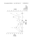 | 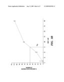 |
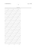 |  |
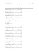 | 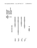 |
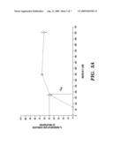 | 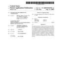 |
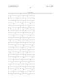 | 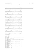 |
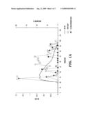 |  |
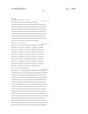 |  |
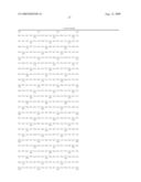 |  |
 |  |
 | 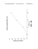 |
| Similar patent applications: | |
| Date | Title |
|---|---|
| 2010-07-22 | Rna interference mediated inhibition of protein tyrosine phosphatase-1b (ptp-1b) gene expression using short interfering nucleic acid (sina) |
| 2010-07-22 | Extended dicer substrate agents and methods for the specific inhibition of gene expression |
| 2010-03-04 | Method for inhibiting angiogenesis |
| 2008-12-18 | Inhibition of tnf-alpha-initiated response |
| 2010-07-08 | Compounds for the inhibition of dgat1 activity |
| New patent applications in this class: | |
| Date | Title |
|---|---|
| 2010-10-21 | Use of cytochrome p450-metabolized drugs and grf molecules in combination therapy |
| 2010-10-21 | Use of protease inhibitors and grf molecules in combination therapy |
| 2010-10-14 | Tissue adhesive using engineered proteins |
| 2010-10-14 | Purification and use of a factor for supporting wound healing |
| 2010-09-30 | Nutritional composition comprising curcuminoids and methods of manufacture |
| New patent applications from these inventors: | |
| Date | Title |
|---|---|
| 2022-09-15 | Large scale production of exosome mimetics and uses thereof |
| 2022-09-15 | Branched poly(-amino esters) for the delivery of nucleic acids |
| 2022-09-08 | Intercellular adhesion molecule 1 (icam1) antibody drug conjugate and uses thereof |
| 2022-09-08 | Components with high api loading |
| 2022-09-01 | Use of rab7 gtpase (rab7) inhibitors in enhancing permeability of the blood brain barrier (bbb) |
| Top Inventors for class "Drug, bio-affecting and body treating compositions" | |
| Rank | Inventor's name |
|---|---|
| 1 | Anthony W. Czarnik |
| 2 | Ulrike Wachendorff-Neumann |
| 3 | Ken Chow |
| 4 | John E. Donello |
| 5 | Rajinder Singh |