Patent application title: METHODS AND COMPOSITIONS FOR DIAGNOSIS AND PROGNOSIS OF RENAL INJURY AND RENAL FAILURE
Inventors:
Joseph Anderberg (Encinitas, CA, US)
Joseph Anderberg (Encinitas, CA, US)
Jeff Gray (Solana Beach, CA, US)
Jeff Gray (Solana Beach, CA, US)
Paul Mcpherson (Encinitas, CA, US)
Paul Mcpherson (Encinitas, CA, US)
Kevin Nakamura (Cardiff By The Sea, CA, US)
Kevin Nakamura (Cardiff By The Sea, CA, US)
Assignees:
ASTUTE MEDICAL, INC.
IPC8 Class: AC12Q137FI
USPC Class:
435 23
Class name: Measuring or testing process involving enzymes or micro-organisms; composition or test strip therefore; processes of forming such composition or test strip involving hydrolase involving proteinase
Publication date: 2011-08-25
Patent application number: 20110207161
Abstract:
The present invention relates to methods and compositions for monitoring,
diagnosis, prognosis, and determination of treatment regimens in subjects
suffering from or suspected of having a renal injury. In particular, the
invention relates to using assays that detect one or more markers
selected from the group consisting of soluble CD44 antigen,
Angiopoietin-1, soluble Angiopoietin-1 receptor, C--X--C chemokine motif
5, soluble Endoglin, soluble Tumor-associated calcium signal transducer
1, Erythropoietin, soluble Fractalkine, Heme oxygenase 1, soluble
Interleukin-1 receptor type II, soluble Interleukin-6 receptor
subunit-alpha, Lymphotactin, Lymphotoxin-alpha, Stromelysin-1, C--C motif
chemokine 22, C--C motif chemokine 5, and Thrombospondin-1 as diagnostic
and prognostic biomarkers in renal injuries.Claims:
1. A method for evaluating renal status in a subject, comprising:
performing one or more assays configured to detect a kidney injury marker
selected from the group consisting of soluble CD44 antigen,
Angiopoietin-1, soluble Angiopoietin-1 receptor, C--X--C chemokine motif
5, soluble Endoglin, soluble Tumor-associated calcium signal transducer
1, Erythropoietin, soluble Fractalkine, Heme oxygenase 1, soluble
Interleukin-1 receptor type II, soluble Interleukin-6 receptor
subunit-alpha, Lymphotactin, Lymphotoxin-alpha, Stromelysin-1, C--C motif
chemokine 22, C--C motif chemokine 5, and Thrombospondin-1 on a body
fluid sample obtained from the subject to provide one or more assay
results; and correlating the assay result(s) to the renal status of the
subject.
2. A method according to claim 1, wherein said correlation step comprises correlating the assay result(s) to one or more of risk stratification, diagnosis, staging, prognosis, classifying and monitoring of the renal status of the subject.
3. A method according to claim 1, wherein said correlating step comprises assigning a likelihood of one or more future changes in renal status to the subject based on the assay result(s).
4. A method according to claim 3, wherein said one or more future changes in renal status comprise one or more of a future injury to renal function, future reduced renal function, future improvement in renal function, and future acute renal failure (ARF).
5. A method according to claim 4, wherein said assay result(s) comprise one or more of: (i) a measured concentration of soluble CD44 antigen, (ii) a measured concentration of Angiopoietin-1, (iii) a measured concentration of soluble Angiopoietin-1 receptor, (iv) a measured concentration of C--X--C chemokine motif 5, (v) a measured concentration of soluble Endoglin, (vi) a measured concentration of soluble Tumor-associated calcium signal transducer 1, (vii) a measured concentration of Erythropoietin, (viii) a measured concentration of soluble Fractalkine, (ix) a measured concentration of Heme oxygenase 1, (x) a measured concentration of soluble Interleukin-1 receptor type II, (xi) a measured concentration of soluble Interleukin-6 receptor subunit-alpha, (xii) a measured concentration of Lymphotactin, (xiii) a measured concentration of Lymphotoxin-alpha, (xiv) a measured concentration of Stromelysin-1, (xv) a measured concentration of C--C motif chemokine 22, (xvi) a measured concentration of C--C motif chemokine 5, or (xvii) a measured concentration of Thrombospondin-1, and said correlation step comprises, for each assay result, comparing said measure concentration to a threshold concentration, and for a positive going marker, assigning an increased likelihood of suffering a future injury to renal function, future reduced renal function, future ARF, or a future improvement in renal function to the subject when the measured concentration is above the threshold, relative to a likelihood assigned when the measured concentration is below the threshold or assigning a decreased likelihood of suffering a future injury to renal function, future reduced renal function, future ARF, or a future improvement in renal function to the subject when the measured concentration is below the threshold, relative to a likelihood assigned when the measured concentration is above the threshold, or for a negative going marker, assigning an increased likelihood of suffering a future injury to renal function, future reduced renal function, future ARF, or a future improvement in renal function to the subject when the measured concentration is below the threshold, relative to a likelihood assigned when the measured concentration is above the threshold or assigning a decreased likelihood of suffering a future injury to renal function, future reduced renal function, future ARF, or a future improvement in renal function to the subject when the measured concentration is above the threshold, relative to a likelihood assigned when the measured concentration is below the threshold.
6. A method according to claim 3, wherein said one or more future changes in renal status comprise a clinical outcome related to a renal injury suffered by the subject.
7. A method according to claim 1, wherein said assay result(s) comprise one or more of: (i) a measured concentration of soluble CD44 antigen, (ii) a measured concentration of Angiopoietin-1, (iii) a measured concentration of soluble Angiopoietin-1 receptor, (iv) a measured concentration of C--X--C chemokine motif 5, (v) a measured concentration of soluble Endoglin, (vi) a measured concentration of soluble Tumor-associated calcium signal transducer 1, (vii) a measured concentration of Erythropoietin, (viii) a measured concentration of soluble Fractalkine, (ix) a measured concentration of Heme oxygenase 1, (x) a measured concentration of soluble Interleukin-1 receptor type II, (xi) a measured concentration of soluble Interleukin-6 receptor subunit-alpha, (xii) a measured concentration of Lymphotactin, (xiii) a measured concentration of Lymphotoxin-alpha, (xiv) a measured concentration of Stromelysin-1, (xv) a measured concentration of C--C motif chemokine 22, (xvi) a measured concentration of C--C motif chemokine 5, or (xvii) a measured concentration of Thrombospondin-1, and said correlation step comprises, for each assay result, comparing said measure concentration to a threshold concentration, and for a positive going marker, assigning an increased likelihood of subsequent acute kidney injury, worsening stage of AKI, mortality, need for renal replacement therapy, need for withdrawal of renal toxins, end stage renal disease, heart failure, stroke, myocardial infarction, or chronic kidney disease to the subject when the measured concentration is above the threshold, relative to a likelihood assigned when the measured concentration is below the threshold, or assigning a decreased likelihood of subsequent acute kidney injury, worsening stage of AKI, mortality, need for renal replacement therapy, need for withdrawal of renal toxins, end stage renal disease, heart failure, stroke, myocardial infarction, or chronic kidney disease to the subject when the measured concentration is below the threshold, relative to a likelihood assigned when the measured concentration is above the threshold, or for a negative going marker, assigning an increased likelihood of subsequent acute kidney injury, worsening stage of AKI, mortality, need for renal replacement therapy, need for withdrawal of renal toxins, end stage renal disease, heart failure, stroke, myocardial infarction, or chronic kidney disease to the subject when the measured concentration is below the threshold, relative to a likelihood assigned when the measured concentration is above the threshold, or assigning a decreased likelihood of subsequent acute kidney injury, worsening stage of AKI, mortality, need for renal replacement therapy, need for withdrawal of renal toxins, end stage renal disease, heart failure, stroke, myocardial infarction, or chronic kidney disease to the subject when the measured concentration is above the threshold, relative to a likelihood assigned when the measured concentration is below the threshold.
8. A method according to claim 3, wherein the likelihood of one or more future changes in renal status is that an event of interest is more or less likely to occur within 30 days of the time at which the body fluid sample is obtained from the subject.
9. A method according to claim 8, wherein the likelihood of one or more future changes in renal status is that an event of interest is more or less likely to occur within a period selected from the group consisting of 21 days, 14 days, 7 days, 5 days, 96 hours, 72 hours, 48 hours, 36 hours, 24 hours, and 12 hours.
10. A method according to claim 1, wherein the subject is selected for evaluation of renal status based on the pre-existence in the subject of one or more known risk factors for prerenal, intrinsic renal, or postrenal ARF.
11. A method according to claim 1, wherein the subject is selected for evaluation of renal status based on an existing diagnosis of one or more of congestive heart failure, preeclampsia, eclampsia, diabetes mellitus, hypertension, coronary artery disease, proteinuria, renal insufficiency, glomerular filtration below the normal range, cirrhosis, serum creatinine above the normal range, sepsis, injury to renal function, reduced renal function, or ARF, or based on undergoing or having undergone major vascular surgery, coronary artery bypass, or other cardiac surgery, or based on exposure to NSAIDs, cyclosporines, tacrolimus, aminoglycosides, foscarnet, ethylene glycol, hemoglobin, myoglobin, ifosfamide, heavy metals, methotrexate, radiopaque contrast agents, or streptozotocin.
12. A method according to claim 1, wherein said correlating step comprises assigning a diagnosis of the occurrence or nonoccurrence of one or more of an injury to renal function, reduced renal function, or ARF to the subject based on the assay result(s).
13. A method according to claim 12, wherein said assay result(s) comprise one or more of: (i) a measured concentration of soluble CD44 antigen, (ii) a measured concentration of Angiopoietin-1, (iii) a measured concentration of soluble Angiopoietin-1 receptor, (iv) a measured concentration of C--X--C chemokine motif 5, (v) a measured concentration of soluble Endoglin, (vi) a measured concentration of soluble Tumor-associated calcium signal transducer 1, (vii) a measured concentration of Erythropoietin, (viii) a measured concentration of soluble Fractalkine, (ix) a measured concentration of Heme oxygenase 1, (x) a measured concentration of soluble Interleukin-1 receptor type II, (xi) a measured concentration of soluble Interleukin-6 receptor subunit-alpha, (xii) a measured concentration of Lymphotactin, (xiii) a measured concentration of Lymphotoxin-alpha, (xiv) a measured concentration of Stromelysin-1, (xv) a measured concentration of C--C motif chemokine 22, (xvi) a measured concentration of C--C motif chemokine 5, or (xvii) a measured concentration of Thrombospondin-1, and said correlation step comprises, for each assay result, comparing said measure concentration to a threshold concentration, and for a positive going marker, assigning the occurrence of an injury to renal function, reduced renal function, or ARF to the subject when the measured concentration is above the threshold, or assigning the nonoccurrence of an injury to renal function, reduced renal function, or ARF to the subject when the measured concentration is below the threshold, or for a negative going marker, assigning the occurrence of an injury to renal function, reduced renal function, or ARF to the subject when the measured concentration is below the threshold, or assigning the nonoccurrence of an injury to renal function, reduced renal function, or ARF to the subject when the measured concentration is above the threshold.
14. A method according to claim 1, wherein said correlating step comprises assessing whether or not renal function is improving or worsening in a subject who has suffered from an injury to renal function, reduced renal function, or ARF based on the assay result(s).
15. A method according to claim 14, wherein said assay result(s) comprise one or more of: (i) a measured concentration of soluble CD44 antigen, (ii) a measured concentration of Angiopoietin-1, (iii) a measured concentration of soluble Angiopoietin-1 receptor, (iv) a measured concentration of C--X--C chemokine motif 5, (v) a measured concentration of soluble Endoglin, (vi) a measured concentration of soluble Tumor-associated calcium signal transducer 1, (vii) a measured concentration of Erythropoietin, (viii) a measured concentration of soluble Fractalkine, (ix) a measured concentration of Heme oxygenase 1, (x) a measured concentration of soluble Interleukin-1 receptor type II, (xi) a measured concentration of soluble Interleukin-6 receptor subunit-alpha, (xii) a measured concentration of Lymphotactin, (xiii) a measured concentration of Lymphotoxin-alpha, (xiv) a measured concentration of Stromelysin-1, (xv) a measured concentration of C--C motif chemokine 22, (xvi) a measured concentration of C--C motif chemokine 5, or (xvii) a measured concentration of Thrombospondin-1, and said correlation step comprises, for each assay result, comparing said measure concentration to a threshold concentration, and for a positive going marker, assigning a worsening of renal function to the subject when the measured concentration is above the threshold, or assigning an improvement of renal function when the measured concentration is below the threshold, or for a negative going marker, assigning a worsening of renal function to the subject when the measured concentration is below the threshold, or assigning an improvement of renal function when the measured concentration is above the threshold.
16. A method according to claim 1, wherein said method is a method of diagnosing the occurrence or nonoccurrence of an injury to renal function in said subject.
17. A method according to claim 1, wherein said method is a method of diagnosing the occurrence or nonoccurrence of reduced renal function in said subject.
18. A method according to claim 1, wherein said method is a method of diagnosing the occurrence or nonoccurrence of acute renal failure in said subject.
19. A method according to claim 1, wherein said method is a method of diagnosing the occurrence or nonoccurrence of a need for renal replacement therapy in said subject.
20. A method according to claim 1, wherein said method is a method of diagnosing the occurrence or nonoccurrence of a need for renal transplantation in said subject.
21. A method according to claim 1, wherein said method is a method of assigning a risk of the future occurrence or nonoccurrence of an injury to renal function in said subject.
22. A method according to claim 1, wherein said method is a method of assigning a risk of the future occurrence or nonoccurrence of reduced renal function in said subject.
23. A method according to claim 1, wherein said method is a method of assigning a risk of the future occurrence or nonoccurrence of acute renal failure in said subject.
24. A method according to claim 1, wherein said method is a method of assigning a risk of the future occurrence or nonoccurrence of a need for renal replacement therapy in said subject.
25. A method according to claim 1, wherein said method is a method of assigning a risk of the future occurrence or nonoccurrence of a need for renal transplantation in said subject.
26. A method according to claim 5, wherein said one or more future changes in renal status comprise one or more of a future injury to renal function, future reduced renal function, future improvement in renal function, and future acute renal failure (ARF) within 72 hours of the time at which the body fluid sample is obtained.
27. A method according to claim 5, wherein said one or more future changes in renal status comprise one or more of a future injury to renal function, future reduced renal function, future improvement in renal function, and future acute renal failure (ARF) within 48 hours of the time at which the body fluid sample is obtained.
28. A method according to claim 5, wherein said one or more future changes in renal status comprise one or more of a future injury to renal function, future reduced renal function, future improvement in renal function, and future acute renal failure (ARF) within 24 hours of the time at which the body fluid sample is obtained.
29. Use of one or more kidney injury markers selected from the group consisting of soluble CD44 antigen, Angiopoietin-1, soluble Angiopoietin-1 receptor, C--X--C chemokine motif 5, soluble Endoglin, soluble Tumor-associated calcium signal transducer 1, Erythropoietin, soluble Fractalkine, Heme oxygenase 1, soluble Interleukin-1 receptor type II, soluble Interleukin-6 receptor subunit-alpha, Lymphotactin, Lymphotoxin-alpha, Stromelysin-1, C--C motif chemokine 22, C--C motif chemokine 5, and Thrombospondin-1 for the evaluation of renal injury.
30. Use of one or more kidney injury markers selected from the group consisting of soluble CD44 antigen, Angiopoietin-1, soluble Angiopoietin-1 receptor, C--X--C chemokine motif 5, soluble Endoglin, soluble Tumor-associated calcium signal transducer 1, Erythropoietin, soluble Fractalkine, Heme oxygenase 1, soluble Interleukin-1 receptor type II, soluble Interleukin-6 receptor subunit-alpha, Lymphotactin, Lymphotoxin-alpha, Stromelysin-1, C--C motif chemokine 22, C--C motif chemokine 5, and Thrombospondin-1 for the evaluation of acute renal injury.
31. A method according to claim 7, wherein the increased or decreased likelihood of subsequent acute kidney injury, worsening stage of AKI, mortality, need for renal replacement therapy, need for withdrawal of renal toxins, end stage renal disease, heart failure, stroke, myocardial infarction, or chronic kidney disease assigned to the subject is a likelihood that an event of interest is more or less likely to occur within 30 days of the time at which the body fluid sample is obtained from the subject.
32. A method according to claim 7, wherein the increased or decreased likelihood of subsequent acute kidney injury, worsening stage of AKI, mortality, need for renal replacement therapy, need for withdrawal of renal toxins, end stage renal disease, heart failure, stroke, myocardial infarction, or chronic kidney disease assigned to the subject is a likelihood that an event of interest is more or less likely to occur within 72 hours of the time at which the body fluid sample is obtained from the subject.
33. A method according to claim 7, wherein the increased or decreased likelihood of subsequent acute kidney injury, worsening stage of AKI, mortality, need for renal replacement therapy, need for withdrawal of renal toxins, end stage renal disease, heart failure, stroke, myocardial infarction, or chronic kidney disease assigned to the subject is a likelihood that an event of interest is more or less likely to occur within 24 hours of the time at which the body fluid sample is obtained from the subject.
Description:
[0001] The present invention claims priority from U.S. Provisional Patent
Application 61/107,287 filed Oct. 21, 2008; U.S. Provisional Patent
Application 61/117,138 filed Nov. 22, 2008; U.S. Provisional Patent
Application 61/113,034 filed Nov. 10, 2008; U.S. Provisional Patent
Application 61/115,050 filed Nov. 15, 2008; U.S. Provisional Patent
Application 61/113,078 filed Nov. 10, 2008; U.S. Provisional Patent
Application 61/113,039 filed Nov. 10, 2008; U.S. Provisional Patent
Application 61/117,143 filed Nov. 22, 2008; U.S. Provisional Patent
Application 61/107,307 filed Oct. 21, 2008; U.S. Provisional Patent
Application 61/113,069 filed Nov. 10, 2008; U.S. Provisional Patent
Application 61/107,293 filed Oct. 21, 2008; U.S. Provisional Patent
Application 61/113,059 filed Nov. 10, 2008; U.S. Provisional Patent
Application 61/115,028 filed Nov. 14, 2008; U.S. Provisional Patent
Application 61/115,026 filed Nov. 14, 2008; U.S. Provisional Patent
Application 61/113,093 filed Nov. 10, 2008; U.S. Provisional Patent
Application 61/115,046 filed Nov. 15, 2008; U.S. Provisional Patent
Application 61/115,043 filed Nov. 15, 2008; and U.S. Provisional Patent
Application 61/117,136 filed Nov. 22, 2008, each of which is hereby
incorporated in its entirety including all tables, figures, and claims.
BACKGROUND OF THE INVENTION
[0002] The following discussion of the background of the invention is merely provided to aid the reader in understanding the invention and is not admitted to describe or constitute prior art to the present invention.
[0003] The kidney is responsible for water and solute excretion from the body. Its functions include maintenance of acid-base balance, regulation of electrolyte concentrations, control of blood volume, and regulation of blood pressure. As such, loss of kidney function through injury and/or disease results in substantial morbidity and mortality. A detailed discussion of renal injuries is provided in Harrison's Principles of Internal Medicine, 17th Ed., McGraw Hill, New York, pages 1741-1830, which are hereby incorporated by reference in their entirety. Renal disease and/or injury may be acute or chronic. Acute and chronic kidney disease are described as follows (from Current Medical Diagnosis & Treatment 2008, 47th Ed, McGraw Hill, New York, pages 785-815, which are hereby incorporated by reference in their entirety): "Acute renal failure is worsening of renal function over hours to days, resulting in the retention of nitrogenous wastes (such as urea nitrogen) and creatinine in the blood. Retention of these substances is called azotemia. Chronic renal failure (chronic kidney disease) results from an abnormal loss of renal function over months to years".
[0004] Acute renal failure (ARF, also known as acute kidney injury, or AKI) is an abrupt (typically detected within about 48 hours to 1 week) reduction in glomerular filtration. This loss of filtration capacity results in retention of nitrogenous (urea and creatinine) and non-nitrogenous waste products that are normally excreted by the kidney, a reduction in urine output, or both. It is reported that ARF complicates about 5% of hospital admissions, 4-15% of cardiopulmonary bypass surgeries, and up to 30% of intensive care admissions. ARF may be categorized as prerenal, intrinsic renal, or postrenal in causation. Intrinsic renal disease can be further divided into glomerular, tubular, interstitial, and vascular abnormalities. Major causes of ARF are described in the following table, which is adapted from the Merck Manual, 17th ed., Chapter 222, and which is hereby incorporated by reference in their entirety:
TABLE-US-00001 Type Risk Factors Prerenal ECF volume depletion Excessive diuresis, hemorrhage, GI losses, loss of intravascular fluid into the extravascular space (due to ascites, peritonitis, pancreatitis, or burns), loss of skin and mucus membranes, renal salt- and water-wasting states Low cardiac output Cardiomyopathy, MI, cardiac tamponade, pulmonary embolism, pulmonary hypertension, positive-pressure mechanical ventilation Low systemic vascular Septic shock, liver failure, antihypertensive drugs resistance Increased renal vascular NSAIDs, cyclosporines, tacrolimus, hypercalcemia, resistance anaphylaxis, anesthetics, renal artery obstruction, renal vein thrombosis, sepsis, hepatorenal syndrome Decreased efferent arteriolar ACE inhibitors or angiotensin II receptor blockers tone (leading to decreased GFR from reduced glomerular transcapillary pressure, especially in patients with bilateral renal artery stenosis) Intrinsic Renal Acute tubular injury Ischemia (prolonged or severe prerenal state): surgery, hemorrhage, arterial or venous obstruction; Toxins: NSAIDs, cyclosporines, tacrolimus, aminoglycosides, foscarnet, ethylene glycol, hemoglobin, myoglobin, ifosfamide, heavy metals, methotrexate, radiopaque contrast agents, streptozotocin Acute glomerulonephritis ANCA-associated: Crescentic glomerulonephritis, polyarteritis nodosa, Wegener's granulomatosis; Anti-GBM glomerulonephritis: Goodpasture's syndrome; Immune- complex: Lupus glomerulonephritis, postinfectious glomerulonephritis, cryoglobulinemic glomerulonephritis Acute tubulointerstitial Drug reaction (eg, β-lactams, NSAIDs, sulfonamides, nephritis ciprofloxacin, thiazide diuretics, furosemide, phenytoin, allopurinol, pyelonephritis, papillary necrosis Acute vascular nephropathy Vasculitis, malignant hypertension, thrombotic microangiopathies, scleroderma, atheroembolism Infiltrative diseases Lymphoma, sarcoidosis, leukemia Postrenal Tubular precipitation Uric acid (tumor lysis), sulfonamides, triamterene, acyclovir, indinavir, methotrexate, ethylene glycol ingestion, myeloma protein, myoglobin Ureteral obstruction Intrinsic: Calculi, clots, sloughed renal tissue, fungus ball, edema, malignancy, congenital defects; Extrinsic: Malignancy, retroperitoneal fibrosis, ureteral trauma during surgery or high impact injury Bladder obstruction Mechanical: Benign prostatic hyperplasia, prostate cancer, bladder cancer, urethral strictures, phimosis, paraphimosis, urethral valves, obstructed indwelling urinary catheter; Neurogenic: Anticholinergic drugs, upper or lower motor neuron lesion
[0005] In the case of ischemic ARF, the course of the disease may be divided into four phases. During an initiation phase, which lasts hours to days, reduced perfusion of the kidney is evolving into injury. Glomerular ultrafiltration reduces, the flow of filtrate is reduced due to debris within the tubules, and back leakage of filtrate through injured epithelium occurs. Renal injury can be mediated during this phase by reperfusion of the kidney. Initiation is followed by an extension phase which is characterized by continued ischemic injury and inflammation and may involve endothelial damage and vascular congestion. During the maintenance phase, lasting from 1 to 2 weeks, renal cell injury occurs, and glomerular filtration and urine output reaches a minimum. A recovery phase can follow in which the renal epithelium is repaired and GFR gradually recovers. Despite this, the survival rate of subjects with ARF may be as low as about 60%.
[0006] Acute kidney injury caused by radiocontrast agents (also called contrast media) and other nephrotoxins such as cyclosporine, antibiotics including aminoglycosides and anticancer drugs such as cisplatin manifests over a period of days to about a week. Contrast induced nephropathy (CIN, which is AKI caused by radiocontrast agents) is thought to be caused by intrarenal vasoconstriction (leading to ischemic injury) and from the generation of reactive oxygen species that are directly toxic to renal tubular epithelial cells. CIN classically presents as an acute (onset within 24-48 h) but reversible (peak 3-5 days, resolution within 1 week) rise in blood urea nitrogen and serum creatinine.
[0007] A commonly reported criteria for defining and detecting AKI is an abrupt (typically within about 2-7 days or within a period of hospitalization) elevation of serum creatinine. Although the use of serum creatinine elevation to define and detect AKI is well established, the magnitude of the serum creatinine elevation and the time over which it is measured to define AKI varies considerably among publications. Traditionally, relatively large increases in serum creatinine such as 100%, 200%, an increase of at least 100% to a value over 2 mg/dL and other definitions were used to define AKI. However, the recent trend has been towards using smaller serum creatinine rises to define AKI. The relationship between serum creatinine rise, AKI and the associated health risks are reviewed in Praught and Shlipak, Curr Opin Nephrol Hypertens 14:265-270, 2005 and Chertow et al, J Am Soc Nephrol 16: 3365-3370, 2005, which, with the references listed therein, are hereby incorporated by reference in their entirety. As described in these publications, acute worsening renal function (AKI) and increased risk of death and other detrimental outcomes are now known to be associated with very small increases in serum creatinine. These increases may be determined as a relative (percent) value or a nominal value. Relative increases in serum creatinine as small as 20% from the pre-injury value have been reported to indicate acutely worsening renal function (AKI) and increased health risk, but the more commonly reported value to define AKI and increased health risk is a relative increase of at least 25%. Nominal increases as small as 0.3 mg/dL, 0.2 mg/dL or even 0.1 mg/dL have been reported to indicate worsening renal function and increased risk of death. Various time periods for the serum creatinine to rise to these threshold values have been used to define AKI, for example, ranging from 2 days, 3 days, 7 days, or a variable period defined as the time the patient is in the hospital or intensive care unit. These studies indicate there is not a particular threshold serum creatinine rise (or time period for the rise) for worsening renal function or AKI, but rather a continuous increase in risk with increasing magnitude of serum creatinine rise.
[0008] One study (Lassnigg et all, J Am Soc Nephrol 15:1597-1605, 2004, hereby incorporated by reference in its entirety) investigated both increases and decreases in serum creatinine. Patients with a mild fall in serum creatinine of -0.1 to -0.3 mg/dL following heart surgery had the lowest mortality rate. Patients with a larger fall in serum creatinine (more than or equal to -0.4 mg/dL) or any increase in serum creatinine had a larger mortality rate. These findings caused the authors to conclude that even very subtle changes in renal function (as detected by small creatinine changes within 48 hours of surgery) seriously effect patient's outcomes. In an effort to reach consensus on a unified classification system for using serum creatinine to define AKI in clinical trials and in clinical practice, Bellomo et al., Crit. Care. 8(4):R204-12, 2004, which is hereby incorporated by reference in its entirety, proposes the following classifications for stratifying AKI patients:
"Risk": serum creatinine increased 1.5 fold from baseline OR urine production of <0.5 ml/kg body weight/hr for 6 hours; "Injury": serum creatinine increased 2.0 fold from baseline OR urine production <0.5 ml/kg/hr for 12 h; "Failure": serum creatinine increased 3.0 fold from baseline OR creatinine>355 μmol/l (with a rise of >44) or urine output below 0.3 ml/kg/hr for 24 h or anuria for at least 12 hours; And included two clinical outcomes: "Loss": persistent need for renal replacement therapy for more than four weeks. "ESRD": end stage renal disease--the need for dialysis for more than 3 months. These criteria are called the RIFLE criteria, which provide a useful clinical tool to classify renal status. As discussed in Kellum, Crit. Care Med. 36: S141-45, 2008 and Ricci et al., Kidney Int. 73, 538-546, 2008, each hereby incorporated by reference in its entirety, the RIFLE criteria provide a uniform definition of AKI which has been validated in numerous studies.
[0009] More recently, Mehta et al., Crit. Care 11:R31 (doi:10.1186.cc5713), 2007, hereby incorporated by reference in its entirety, proposes the following similar classifications for stratifying AKI patients, which have been modified from RIFLE:
"Stage I": increase in serum creatinine of more than or equal to 0.3 mg/dL (>26.4 μmol/L) or increase to more than or equal to 150% (1.5-fold) from baseline OR urine output less than 0.5 mL/kg per hour for more than 6 hours; "Stage II": increase in serum creatinine to more than 200% (>26.4 μmol/L) from baseline OR urine output less than 0.5 mL/kg per hour for more than 12 hours; "Stage III": increase in serum creatinine to more than 300% (>3-fold) from baseline OR serum creatinine≧354 μmol/L accompanied by an acute increase of at least 44 μmol/L OR urine output less than 0.3 mL/kg per hour for 24 hours or anuria for 12 hours.
[0010] The CIN Consensus Working Panel (McCollough et al, Rev Cardiovasc Med. 2006; 7(4):177-197, hereby incorporated by reference in its entirety) uses a serum creatinine rise of 25% to define Contrast induced nephropathy (which is a type of AKI). Although various groups propose slightly different criteria for using serum creatinine to detect AKI, the consensus is that small changes in serum creatinine, such as 0.3 mg/dL or 25%, are sufficient to detect AKI (worsening renal function) and that the magnitude of the serum creatinine change is an indicator of the severity of the AKI and mortality risk.
[0011] Although serial measurement of serum creatinine over a period of days is an accepted method of detecting and diagnosing AKI and is considered one of the most important tools to evaluate AKI patients, serum creatinine is generally regarded to have several limitations in the diagnosis, assessment and monitoring of AKI patients. The time period for serum creatinine to rise to values (e.g., a 0.3 mg/dL or 25% rise) considered diagnostic for AKI can be 48 hours or longer depending on the definition used. Since cellular injury in AKI can occur over a period of hours, serum creatinine elevations detected at 48 hours or longer can be a late indicator of injury, and relying on serum creatinine can thus delay diagnosis of AKI. Furthermore, serum creatinine is not a good indicator of the exact kidney status and treatment needs during the most acute phases of AKI when kidney function is changing rapidly. Some patients with AKI will recover fully, some will need dialysis (either short term or long term) and some will have other detrimental outcomes including death, major adverse cardiac events and chronic kidney disease. Because serum creatinine is a marker of filtration rate, it does not differentiate between the causes of AKI (pre-renal, intrinsic renal, post-renal obstruction, atheroembolic, etc) or the category or location of injury in intrinsic renal disease (for example, tubular, glomerular or interstitial in origin). Urine output is similarly limited, Knowing these things can be of vital importance in managing and treating patients with AKI.
[0012] These limitations underscore the need for better methods to detect and assess AKI, particularly in the early and subclinical stages, but also in later stages when recovery and repair of the kidney can occur. Furthermore, there is a need to better identify patients who are at risk of having an AKI.
BRIEF SUMMARY OF THE INVENTION
[0013] It is an object of the invention to provide methods and compositions for evaluating renal function in a subject. As described herein, measurement of one or more markers selected from the group consisting of soluble CD44 antigen, Angiopoietin-1, soluble Angiopoietin-1 receptor, C--X--C chemokine motif 5, soluble Endoglin, soluble Tumor-associated calcium signal transducer 1, Erythropoietin, soluble Fractalkine, Heme oxygenase 1, soluble Interleukin-1 receptor type II, soluble Interleukin-6 receptor subunit-alpha, Lymphotactin, Lymphotoxin-alpha, Stromelysin-1, C--C motif chemokine 22, C--C motif chemokine 5, and Thrombospondin-1 (collectively referred to herein as "kidney injury markers, and individually as a "kidney injury marker") can be used for diagnosis, prognosis, risk stratification, staging, monitoring, categorizing and determination of further diagnosis and treatment regimens in subjects suffering or at risk of suffering from an injury to renal function, reduced renal function, and/or acute renal failure (also called acute kidney injury).
[0014] These kidney injury markers may be used, individually or in panels comprising a plurality of kidney injury markers, for risk stratification (that is, to identify subjects at risk for a future injury to renal function, for future progression to reduced renal function, for future progression to ARF, for future improvement in renal function, etc.); for diagnosis of existing disease (that is, to identify subjects who have suffered an injury to renal function, who have progressed to reduced renal function, who have progressed to ARF, etc.); for monitoring for deterioration or improvement of renal function; and for predicting a future medical outcome, such as improved or worsening renal function, a decreased or increased mortality risk, a decreased or increased risk that a subject will require renal replacement therapy (i.e., hemodialysis, peritoneal dialysis, hemofiltration, and/or renal transplantation, a decreased or increased risk that a subject will recover from an injury to renal function, a decreased or increased risk that a subject will recover from ARF, a decreased or increased risk that a subject will progress to end stage renal disease, a decreased or increased risk that a subject will progress to chronic renal failure, a decreased or increased risk that a subject will suffer rejection of a transplanted kidney, etc.
[0015] In a first aspect, the present invention relates to methods for evaluating renal status in a subject. These methods comprise performing an assay method that is configured to detect one or more kidney injury markers of the present invention in a body fluid sample obtained from the subject. The assay result(s), for example a measured concentration of one or more markers selected from the group consisting of soluble CD44 antigen, Angiopoietin-1, soluble Angiopoietin-1 receptor, C--X--C chemokine motif 5, soluble Endoglin, soluble Tumor-associated calcium signal transducer 1, Erythropoietin, soluble Fractalkine, Heme oxygenase 1, soluble Interleukin-1 receptor type II, soluble Interleukin-6 receptor subunit-alpha, Lymphotactin, Lymphotoxin-alpha, Stromelysin-1, C--C motif chemokine 22, C--C motif chemokine 5, and Thrombospondin-1 is/are then correlated to the renal status of the subject. This correlation to renal status may include correlating the assay result(s) to one or more of risk stratification, diagnosis, prognosis, staging, classifying and monitoring of the subject as described herein. Thus, the present invention utilizes one or more kidney injury markers of the present invention for the evaluation of renal injury.
[0016] In certain embodiments, the methods for evaluating renal status described herein are methods for risk stratification of the subject; that is, assigning a likelihood of one or more future changes in renal status to the subject. In these embodiments, the assay result(s) is/are correlated to one or more such future changes. The following are preferred risk stratification embodiments.
[0017] In preferred risk stratification embodiments, these methods comprise determining a subject's risk for a future injury to renal function, and the assay result(s) is/are correlated to a likelihood of such a future injury to renal function. For example, the measured concentration(s) may each be compared to a threshold value. For a "positive going" kidney injury marker, an increased likelihood of suffering a future injury to renal function is assigned to the subject when the measured concentration is above the threshold, relative to a likelihood assigned when the measured concentration is below the threshold. For a "negative going" kidney injury marker, an increased likelihood of suffering a future injury to renal function is assigned to the subject when the measured concentration is below the threshold, relative to a likelihood assigned when the measured concentration is above the threshold.
[0018] In other preferred risk stratification embodiments, these methods comprise determining a subject's risk for future reduced renal function, and the assay result(s) is/are correlated to a likelihood of such reduced renal function. For example, the measured concentrations may each be compared to a threshold value. For a "positive going" kidney injury marker, an increased likelihood of suffering a future reduced renal function is assigned to the subject when the measured concentration is above the threshold, relative to a likelihood assigned when the measured concentration is below the threshold. For a "negative going" kidney injury marker, an increased likelihood of future reduced renal function is assigned to the subject when the measured concentration is below the threshold, relative to a likelihood assigned when the measured concentration is above the threshold.
[0019] In still other preferred risk stratification embodiments, these methods comprise determining a subject's likelihood for a future improvement in renal function, and the assay result(s) is/are correlated to a likelihood of such a future improvement in renal function. For example, the measured concentration(s) may each be compared to a threshold value. For a "positive going" kidney injury marker, an increased likelihood of a future improvement in renal function is assigned to the subject when the measured concentration is below the threshold, relative to a likelihood assigned when the measured concentration is above the threshold. For a "negative going" kidney injury marker, an increased likelihood of a future improvement in renal function is assigned to the subject when the measured concentration is above the threshold, relative to a likelihood assigned when the measured concentration is below the threshold.
[0020] In yet other preferred risk stratification embodiments, these methods comprise determining a subject's risk for progression to ARF, and the result(s) is/are correlated to a likelihood of such progression to ARF. For example, the measured concentration(s) may each be compared to a threshold value. For a "positive going" kidney injury marker, an increased likelihood of progression to ARF is assigned to the subject when the measured concentration is above the threshold, relative to a likelihood assigned when the measured concentration is below the threshold. For a "negative going" kidney injury marker, an increased likelihood of progression to ARF is assigned to the subject when the measured concentration is below the threshold, relative to a likelihood assigned when the measured concentration is above the threshold.
[0021] And in other preferred risk stratification embodiments, these methods comprise determining a subject's outcome risk, and the assay result(s) is/are correlated to a likelihood of the occurrence of a clinical outcome related to a renal injury suffered by the subject. For example, the measured concentration(s) may each be compared to a threshold value. For a "positive going" kidney injury marker, an increased likelihood of one or more of: acute kidney injury, progression to a worsening stage of AKI, mortality, a requirement for renal replacement therapy, a requirement for withdrawal of renal toxins, end stage renal disease, heart failure, stroke, myocardial infarction, progression to chronic kidney disease, etc., is assigned to the subject when the measured concentration is above the threshold, relative to a likelihood assigned when the measured concentration is below the threshold. For a "negative going" kidney injury marker, an increased likelihood of one or more of: acute kidney injury, progression to a worsening stage of AKI, mortality, a requirement for renal replacement therapy, a requirement for withdrawal of renal toxins, end stage renal disease, heart failure, stroke, myocardial infarction, progression to chronic kidney disease, etc., is assigned to the subject when the measured concentration is below the threshold, relative to a likelihood assigned when the measured concentration is above the threshold.
[0022] In such risk stratification embodiments, preferably the likelihood or risk assigned is that an event of interest is more or less likely to occur within 180 days of the time at which the body fluid sample is obtained from the subject. In particularly preferred embodiments, the likelihood or risk assigned relates to an event of interest occurring within a shorter time period such as 18 months, 120 days, 90 days, 60 days, 45 days, 30 days, 21 days, 14 days, 7 days, 5 days, 96 hours, 72 hours, 48 hours, 36 hours, 24 hours, 12 hours, or less. A risk at 0 hours of the time at which the body fluid sample is obtained from the subject is equivalent to diagnosis of a current condition.
[0023] In preferred risk stratification embodiments, the subject is selected for risk stratification based on the pre-existence in the subject of one or more known risk factors for prerenal, intrinsic renal, or postrenal ARF. For example, a subject undergoing or having undergone major vascular surgery, coronary artery bypass, or other cardiac surgery; a subject having pre-existing congestive heart failure, preeclampsia, eclampsia, diabetes mellitus, hypertension, coronary artery disease, proteinuria, renal insufficiency, glomerular filtration below the normal range, cirrhosis, serum creatinine above the normal range, or sepsis; or a subject exposed to NSAIDs, cyclosporines, tacrolimus, aminoglycosides, foscarnet, ethylene glycol, hemoglobin, myoglobin, ifosfamide, heavy metals, methotrexate, radiopaque contrast agents, or streptozotocin are all preferred subjects for monitoring risks according to the methods described herein. This list is not meant to be limiting. By "pre-existence" in this context is meant that the risk factor exists at the time the body fluid sample is obtained from the subject. In particularly preferred embodiments, a subject is chosen for risk stratification based on an existing diagnosis of injury to renal function, reduced renal function, or ARF.
[0024] In other embodiments, the methods for evaluating renal status described herein are methods for diagnosing a renal injury in the subject; that is, assessing whether or not a subject has suffered from an injury to renal function, reduced renal function, or ARF. In these embodiments, the assay result(s), for example a measured concentration of one or more markers selected from the group consisting of soluble CD44 antigen, Angiopoietin-1, soluble Angiopoietin-1 receptor, C--X--C chemokine motif 5, soluble Endoglin, soluble Tumor-associated calcium signal transducer 1, Erythropoietin, soluble Fractalkine, Heme oxygenase 1, soluble Interleukin-1 receptor type II, soluble Interleukin-6 receptor subunit-alpha, Lymphotactin, Lymphotoxin-alpha, Stromelysin-1, C--C motif chemokine 22, C--C motif chemokine 5, and Thrombospondin-1 is/are correlated to the occurrence or nonoccurrence of a change in renal status. The following are preferred diagnostic embodiments.
[0025] In preferred diagnostic embodiments, these methods comprise diagnosing the occurrence or nonoccurrence of an injury to renal function, and the assay result(s) is/are correlated to the occurrence or nonoccurrence of such an injury. For example, each of the measured concentration(s) may be compared to a threshold value. For a positive going marker, an increased likelihood of the occurrence of an injury to renal function is assigned to the subject when the measured concentration is above the threshold (relative to the likelihood assigned when the measured concentration is below the threshold); alternatively, when the measured concentration is below the threshold, an increased likelihood of the nonoccurrence of an injury to renal function may be assigned to the subject (relative to the likelihood assigned when the measured concentration is above the threshold). For a negative going marker, an increased likelihood of the occurrence of an injury to renal function is assigned to the subject when the measured concentration is below the threshold (relative to the likelihood assigned when the measured concentration is above the threshold); alternatively, when the measured concentration is above the threshold, an increased likelihood of the nonoccurrence of an injury to renal function may be assigned to the subject (relative to the likelihood assigned when the measured concentration is below the threshold).
[0026] In other preferred diagnostic embodiments, these methods comprise diagnosing the occurrence or nonoccurrence of reduced renal function, and the assay result(s) is/are correlated to the occurrence or nonoccurrence of an injury causing reduced renal function. For example, each of the measured concentration(s) may be compared to a threshold value. For a positive going marker, an increased likelihood of the occurrence of an injury causing reduced renal function is assigned to the subject when the measured concentration is above the threshold (relative to the likelihood assigned when the measured concentration is below the threshold); alternatively, when the measured concentration is below the threshold, an increased likelihood of the nonoccurrence of an injury causing reduced renal function may be assigned to the subject (relative to the likelihood assigned when the measured concentration is above the threshold). For a negative going marker, an increased likelihood of the occurrence of an injury causing reduced renal function is assigned to the subject when the measured concentration is below the threshold (relative to the likelihood assigned when the measured concentration is above the threshold); alternatively, when the measured concentration is above the threshold, an increased likelihood of the nonoccurrence of an injury causing reduced renal function may be assigned to the subject (relative to the likelihood assigned when the measured concentration is below the threshold).
[0027] In yet other preferred diagnostic embodiments, these methods comprise diagnosing the occurrence or nonoccurrence of ARF, and the assay result(s) is/are correlated to the occurrence or nonoccurrence of an injury causing ARF. For example, each of the measured concentration(s) may be compared to a threshold value. For a positive going marker, an increased likelihood of the occurrence of ARF is assigned to the subject when the measured concentration is above the threshold (relative to the likelihood assigned when the measured concentration is below the threshold); alternatively, when the measured concentration is below the threshold, an increased likelihood of the nonoccurrence of ARF may be assigned to the subject (relative to the likelihood assigned when the measured concentration is above the threshold). For a negative going marker, an increased likelihood of the occurrence of ARF is assigned to the subject when the measured concentration is below the threshold (relative to the likelihood assigned when the measured concentration is above the threshold); alternatively, when the measured concentration is above the threshold, an increased likelihood of the nonoccurrence of ARF may be assigned to the subject (relative to the likelihood assigned when the measured concentration is below the threshold).
[0028] In still other preferred diagnostic embodiments, these methods comprise diagnosing a subject as being in need of renal replacement therapy, and the assay result(s) is/are correlated to a need for renal replacement therapy. For example, each of the measured concentration(s) may be compared to a threshold value. For a positive going marker, an increased likelihood of the occurrence of an injury creating a need for renal replacement therapy is assigned to the subject when the measured concentration is above the threshold (relative to the likelihood assigned when the measured concentration is below the threshold); alternatively, when the measured concentration is below the threshold, an increased likelihood of the nonoccurrence of an injury creating a need for renal replacement therapy may be assigned to the subject (relative to the likelihood assigned when the measured concentration is above the threshold). For a negative going marker, an increased likelihood of the occurrence of an injury creating a need for renal replacement therapy is assigned to the subject when the measured concentration is below the threshold (relative to the likelihood assigned when the measured concentration is above the threshold); alternatively, when the measured concentration is above the threshold, an increased likelihood of the nonoccurrence of an injury creating a need for renal replacement therapy may be assigned to the subject (relative to the likelihood assigned when the measured concentration is below the threshold).
[0029] In still other preferred diagnostic embodiments, these methods comprise diagnosing a subject as being in need of renal transplantation, and the assay result(s) is/are correlated to a need for renal transplantation. For example, each of the measured concentration(s) may be compared to a threshold value. For a positive going marker, an increased likelihood of the occurrence of an injury creating a need for renal transplantation is assigned to the subject when the measured concentration is above the threshold (relative to the likelihood assigned when the measured concentration is below the threshold); alternatively, when the measured concentration is below the threshold, an increased likelihood of the nonoccurrence of an injury creating a need for renal transplantation may be assigned to the subject (relative to the likelihood assigned when the measured concentration is above the threshold). For a negative going marker, an increased likelihood of the occurrence of an injury creating a need for renal transplantation is assigned to the subject when the measured concentration is below the threshold (relative to the likelihood assigned when the measured concentration is above the threshold); alternatively, when the measured concentration is above the threshold, an increased likelihood of the nonoccurrence of an injury creating a need for renal transplantation may be assigned to the subject (relative to the likelihood assigned when the measured concentration is below the threshold).
[0030] In still other embodiments, the methods for evaluating renal status described herein are methods for monitoring a renal injury in the subject; that is, assessing whether or not renal function is improving or worsening in a subject who has suffered from an injury to renal function, reduced renal function, or ARF. In these embodiments, the assay result(s), for example a measured concentration of one or more markers selected from the group consisting of soluble CD44 antigen, Angiopoietin-1, soluble Angiopoietin-1 receptor, C--X--C chemokine motif 5, soluble Endoglin, soluble Tumor-associated calcium signal transducer 1, Erythropoietin, soluble Fractalkine, Heme oxygenase 1, soluble Interleukin-1 receptor type II, soluble Interleukin-6 receptor subunit-alpha, Lymphotactin, Lymphotoxin-alpha, Stromelysin-1, C--C motif chemokine 22, C--C motif chemokine 5, and Thrombospondin-1 is/are correlated to the occurrence or nonoccurrence of a change in renal status. The following are preferred monitoring embodiments.
[0031] In preferred monitoring embodiments, these methods comprise monitoring renal status in a subject suffering from an injury to renal function, and the assay result(s) is/are correlated to the occurrence or nonoccurrence of a change in renal status in the subject. For example, the measured concentration(s) may be compared to a threshold value. For a positive going marker, when the measured concentration is above the threshold, a worsening of renal function may be assigned to the subject; alternatively, when the measured concentration is below the threshold, an improvement of renal function may be assigned to the subject. For a negative going marker, when the measured concentration is below the threshold, a worsening of renal function may be assigned to the subject; alternatively, when the measured concentration is above the threshold, an improvement of renal function may be assigned to the subject.
[0032] In other preferred monitoring embodiments, these methods comprise monitoring renal status in a subject suffering from reduced renal function, and the assay result(s) is/are correlated to the occurrence or nonoccurrence of a change in renal status in the subject. For example, the measured concentration(s) may be compared to a threshold value. For a positive going marker, when the measured concentration is above the threshold, a worsening of renal function may be assigned to the subject; alternatively, when the measured concentration is below the threshold, an improvement of renal function may be assigned to the subject. For a negative going marker, when the measured concentration is below the threshold, a worsening of renal function may be assigned to the subject; alternatively, when the measured concentration is above the threshold, an improvement of renal function may be assigned to the subject.
[0033] In yet other preferred monitoring embodiments, these methods comprise monitoring renal status in a subject suffering from acute renal failure, and the assay result(s) is/are correlated to the occurrence or nonoccurrence of a change in renal status in the subject. For example, the measured concentration(s) may be compared to a threshold value. For a positive going marker, when the measured concentration is above the threshold, a worsening of renal function may be assigned to the subject; alternatively, when the measured concentration is below the threshold, an improvement of renal function may be assigned to the subject. For a negative going marker, when the measured concentration is below the threshold, a worsening of renal function may be assigned to the subject; alternatively, when the measured concentration is above the threshold, an improvement of renal function may be assigned to the subject.
[0034] In other additional preferred monitoring embodiments, these methods comprise monitoring renal status in a subject at risk of an injury to renal function due to the pre-existence of one or more known risk factors for prerenal, intrinsic renal, or postrenal ARF, and the assay result(s) is/are correlated to the occurrence or nonoccurrence of a change in renal status in the subject. For example, the measured concentration(s) may be compared to a threshold value. For a positive going marker, when the measured concentration is above the threshold, a worsening of renal function may be assigned to the subject; alternatively, when the measured concentration is below the threshold, an improvement of renal function may be assigned to the subject. For a negative going marker, when the measured concentration is below the threshold, a worsening of renal function may be assigned to the subject; alternatively, when the measured concentration is above the threshold, an improvement of renal function may be assigned to the subject.
[0035] In still other embodiments, the methods for evaluating renal status described herein are methods for classifying a renal injury in the subject; that is, determining whether a renal injury in a subject is prerenal, intrinsic renal, or postrenal; and/or further subdividing these classes into subclasses such as acute tubular injury, acute glomerulonephritis acute tubulointerstitial nephritis, acute vascular nephropathy, or infiltrative disease; and/or assigning a likelihood that a subject will progress to a particular RIFLE stage. In these embodiments, the assay result(s), for example a measured concentration of one or more markers selected from the group consisting of soluble CD44 antigen, Angiopoietin-1, soluble Angiopoietin-1 receptor, C--X--C chemokine motif 5, soluble Endoglin, soluble Tumor-associated calcium signal transducer 1, Erythropoietin, soluble Fractalkine, Heme oxygenase 1, soluble Interleukin-1 receptor type II, soluble Interleukin-6 receptor subunit-alpha, Lymphotactin, Lymphotoxin-alpha, Stromelysin-1, C--C motif chemokine 22, C--C motif chemokine 5, and Thrombospondin-1 is/are correlated to a particular class and/or subclass. The following are preferred classification embodiments.
[0036] In preferred classification embodiments, these methods comprise determining whether a renal injury in a subject is prerenal, intrinsic renal, or postrenal; and/or further subdividing these classes into subclasses such as acute tubular injury, acute glomerulonephritis acute tubulointerstitial nephritis, acute vascular nephropathy, or infiltrative disease; and/or assigning a likelihood that a subject will progress to a particular RIFLE stage, and the assay result(s) is/are correlated to the injury classification for the subject. For example, the measured concentration may be compared to a threshold value, and when the measured concentration is above the threshold, a particular classification is assigned; alternatively, when the measured concentration is below the threshold, a different classification may be assigned to the subject.
[0037] A variety of methods may be used by the skilled artisan to arrive at a desired threshold value for use in these methods. For example, the threshold value may be determined from a population of normal subjects by selecting a concentration representing the 75th, 85th, 90th, 95th, or 99th percentile of a kidney injury marker measured in such normal subjects. Alternatively, the threshold value may be determined from a "diseased" population of subjects, e.g., those suffering from an injury or having a predisposition for an injury (e.g., progression to ARF or some other clinical outcome such as death, dialysis, renal transplantation, etc.), by selecting a concentration representing the 75th, 85th, 90th, 95th, or 99th percentile of a kidney injury marker measured in such subjects. In another alternative, the threshold value may be determined from a prior measurement of a kidney injury marker in the same subject; that is, a temporal change in the level of a kidney injury marker in the subject may be used to assign risk to the subject.
[0038] The foregoing discussion is not meant to imply, however, that the kidney injury markers of the present invention must be compared to corresponding individual thresholds. Methods for combining assay results can comprise the use of multivariate logistical regression, loglinear modeling, neural network analysis, n-of-m analysis, decision tree analysis, calculating ratios of markers, etc. This list is not meant to be limiting. In these methods, a composite result which is determined by combining individual markers may be treated as if it is itself a marker; that is, a threshold may be determined for the composite result as described herein for individual markers, and the composite result for an individual patient compared to this threshold.
[0039] The ability of a particular test to distinguish two populations can be established using ROC analysis. For example, ROC curves established from a "first" subpopulation which is predisposed to one or more future changes in renal status, and a "second" subpopulation which is not so predisposed can be used to calculate a ROC curve, and the area under the curve provides a measure of the quality of the test. Preferably, the tests described herein provide a ROC curve area greater than 0.5, preferably at least 0.6, more preferably 0.7, still more preferably at least 0.8, even more preferably at least 0.9, and most preferably at least 0.95.
[0040] In certain aspects, the measured concentration of one or more kidney injury markers, or a composite of such markers, may be treated as continuous variables. For example, any particular concentration can be converted into a corresponding probability of a future reduction in renal function for the subject, the occurrence of an injury, a classification, etc. In yet another alternative, a threshold that can provide an acceptable level of specificity and sensitivity in separating a population of subjects into "bins" such as a "first" subpopulation (e.g., which is predisposed to one or more future changes in renal status, the occurrence of an injury, a classification, etc.) and a "second" subpopulation which is not so predisposed. A threshold value is selected to separate this first and second population by one or more of the following measures of test accuracy:
an odds ratio greater than 1, preferably at least about 2 or more or about 0.5 or less, more preferably at least about 3 or more or about 0.33 or less, still more preferably at least about 4 or more or about 0.25 or less, even more preferably at least about 5 or more or about 0.2 or less, and most preferably at least about 10 or more or about 0.1 or less; a specificity of greater than 0.5, preferably at least about 0.6, more preferably at least about 0.7, still more preferably at least about 0.8, even more preferably at least about 0.9 and most preferably at least about 0.95, with a corresponding sensitivity greater than 0.2, preferably greater than about 0.3, more preferably greater than about 0.4, still more preferably at least about 0.5, even more preferably about 0.6, yet more preferably greater than about 0.7, still more preferably greater than about 0.8, more preferably greater than about 0.9, and most preferably greater than about 0.95; a sensitivity of greater than 0.5, preferably at least about 0.6, more preferably at least about 0.7, still more preferably at least about 0.8, even more preferably at least about 0.9 and most preferably at least about 0.95, with a corresponding specificity greater than 0.2, preferably greater than about 0.3, more preferably greater than about 0.4, still more preferably at least about 0.5, even more preferably about 0.6, yet more preferably greater than about 0.7, still more preferably greater than about 0.8, more preferably greater than about 0.9, and most preferably greater than about 0.95; at least about 75% sensitivity, combined with at least about 75% specificity; a positive likelihood ratio (calculated as sensitivity/(1-specificity)) of greater than 1, at least about 2, more preferably at least about 3, still more preferably at least about 5, and most preferably at least about 10; or a negative likelihood ratio (calculated as (1-sensitivity)/specificity) of less than 1, less than or equal to about 0.5, more preferably less than or equal to about 0.3, and most preferably less than or equal to about 0.1. The term "about" in the context of any of the above measurements refers to +/-5% of a given measurement.
[0041] Multiple thresholds may also be used to assess renal status in a subject. For example, a "first" subpopulation which is predisposed to one or more future changes in renal status, the occurrence of an injury, a classification, etc., and a "second" subpopulation which is not so predisposed can be combined into a single group. This group is then subdivided into three or more equal parts (known as tertiles, quartiles, quintiles, etc., depending on the number of subdivisions). An odds ratio is assigned to subjects based on which subdivision they fall into. If one considers a tertile, the lowest or highest tertile can be used as a reference for comparison of the other subdivisions. This reference subdivision is assigned an odds ratio of 1. The second tertile is assigned an odds ratio that is relative to that first tertile. That is, someone in the second tertile might be 3 times more likely to suffer one or more future changes in renal status in comparison to someone in the first tertile. The third tertile is also assigned an odds ratio that is relative to that first tertile.
[0042] In certain embodiments, the assay method is an immunoassay. Antibodies for use in such assays will specifically bind a full length kidney injury marker of interest, and may also bind one or more polypeptides that are "related" thereto, as that term is defined hereinafter. Numerous immunoassay formats are known to those of skill in the art. Preferred body fluid samples are selected from the group consisting of urine, blood, serum, saliva, tears, and plasma.
[0043] The foregoing method steps should not be interpreted to mean that the kidney injury marker assay result(s) is/are used in isolation in the methods described herein. Rather, additional variables or other clinical indicia may be included in the methods described herein. For example, a risk stratification, diagnostic, classification, monitoring, etc. method may combine the assay result(s) with one or more variables measured for the subject selected from the group consisting of demographic information (e.g., weight, sex, age, race), medical history (e.g., family history, type of surgery, pre-existing disease such as aneurism, congestive heart failure, preeclampsia, eclampsia, diabetes mellitus, hypertension, coronary artery disease, proteinuria, renal insufficiency, or sepsis, type of toxin exposure such as NSAIDs, cyclosporines, tacrolimus, aminoglycosides, foscarnet, ethylene glycol, hemoglobin, myoglobin, ifosfamide, heavy metals, methotrexate, radiopaque contrast agents, or streptozotocin), clinical variables (e.g., blood pressure, temperature, respiration rate), risk scores (APACHE score, PREDICT score, TIMI Risk Score for UA/NSTEMI, Framingham Risk Score), a glomerular filtration rate, an estimated glomerular filtration rate, a urine production rate, a serum or plasma creatinine concentration, a urine creatinine concentration, a fractional excretion of sodium, a urine sodium concentration, a urine creatinine to serum or plasma creatinine ratio, a urine specific gravity, a urine osmolality, a urine urea nitrogen to plasma urea nitrogen ratio, a plasma BUN to creatinine ratio, a renal failure index calculated as urine sodium/(urine creatinine/plasma creatinine), a serum or plasma neutrophil gelatinase (NGAL) concentration, a urine NGAL concentration, a serum or plasma cystatin C concentration, a serum or plasma cardiac troponin concentration, a serum or plasma BNP concentration, a serum or plasma NTproBNP concentration, and a serum or plasma proBNP concentration. Other measures of renal function which may be combined with one or more kidney injury marker assay result(s) are described hereinafter and in Harrison's Principles of Internal Medicine, 17th Ed., McGraw Hill, New York, pages 1741-1830, and Current Medical Diagnosis & Treatment 2008, 47th Ed, McGraw Hill, New York, pages 785-815, each of which are hereby incorporated by reference in their entirety.
[0044] When more than one marker is measured, the individual markers may be measured in samples obtained at the same time, or may be determined from samples obtained at different (e.g., an earlier or later) times. The individual markers may also be measured on the same or different body fluid samples. For example, one kidney injury marker may be measured in a serum or plasma sample and another kidney injury marker may be measured in a urine sample. In addition, assignment of a likelihood may combine an individual kidney injury marker assay result with temporal changes in one or more additional variables.
[0045] In various related aspects, the present invention also relates to devices and kits for performing the methods described herein. Suitable kits comprise reagents sufficient for performing an assay for at least one of the described kidney injury markers, together with instructions for performing the described threshold comparisons.
[0046] In certain embodiments, reagents for performing such assays are provided in an assay device, and such assay devices may be included in such a kit. Preferred reagents can comprise one or more solid phase antibodies, the solid phase antibody comprising antibody that detects the intended biomarker target(s) bound to a solid support. In the case of sandwich immunoassays, such reagents can also include one or more detectably labeled antibodies, the detectably labeled antibody comprising antibody that detects the intended biomarker target(s) bound to a detectable label. Additional optional elements that may be provided as part of an assay device are described hereinafter.
[0047] Detectable labels may include molecules that are themselves detectable (e.g., fluorescent moieties, electrochemical labels, ecl (electrochemical luminescence) labels, metal chelates, colloidal metal particles, etc.) as well as molecules that may be indirectly detected by production of a detectable reaction product (e.g., enzymes such as horseradish peroxidase, alkaline phosphatase, etc.) or through the use of a specific binding molecule which itself may be detectable (e.g., a labeled antibody that binds to the second antibody, biotin, digoxigenin, maltose, oligohistidine, 2,4-dintrobenzene, phenylarsenate, ssDNA, dsDNA, etc.).
[0048] Generation of a signal from the signal development element can be performed using various optical, acoustical, and electrochemical methods well known in the art. Examples of detection modes include fluorescence, radiochemical detection, reflectance, absorbance, amperometry, conductance, impedance, interferometry, ellipsometry, etc. In certain of these methods, the solid phase antibody is coupled to a transducer (e.g., a diffraction grating, electrochemical sensor, etc) for generation of a signal, while in others, a signal is generated by a transducer that is spatially separate from the solid phase antibody (e.g., a fluorometer that employs an excitation light source and an optical detector). This list is not meant to be limiting. Antibody-based biosensors may also be employed to determine the presence or amount of analytes that optionally eliminate the need for a labeled molecule.
BRIEF DESCRIPTION OF THE FIGURES
[0049] FIG. 1 provides data tables determined in accordance with Example 6 for the comparison of marker levels in urine samples collected for Cohort 1 (patients that did not progress beyond RIFLE stage 0) and in urine samples collected from subjects at 0, 24 hours, and 48 hours prior to reaching stage R, I or F in Cohort 2. Tables provide descriptive statistics, AUC analysis, and sensitivity, specificity and odds ratio calculations at various threshold (cutoff) levels for the various markers.
[0050] FIG. 2 provides data tables determined in accordance with Example 7 for the comparison of marker levels in urine samples collected for Cohort 1 (patients that did not progress beyond RIFLE stage 0 or R) and in urine samples collected from subjects at 0, 24 hours, and 48 hours prior to reaching stage I or F in Cohort 2. Tables provide descriptive statistics, AUC analysis, and sensitivity, specificity and odds ratio calculations at various threshold (cutoff) levels for the various markers.
[0051] FIG. 3 provides data tables determined in accordance with Example 8 for the comparison of marker levels in urine samples collected for Cohort 1 (patients that reached, but did not progress beyond, RIFLE stage R) and in urine samples collected from subjects at 0, 24 hours, and 48 hours prior to reaching stage I or F in Cohort 2. Tables provide descriptive statistics, AUC analysis, and sensitivity, specificity and odds ratio calculations at various threshold (cutoff) levels for the various markers.
[0052] FIG. 4 provides data tables determined in accordance with Example 9 for the comparison of marker levels in urine samples collected for Cohort 1 (patients that did not progress beyond RIFLE stage 0) and in urine samples collected from subjects at 0, 24 hours, and 48 hours prior to reaching stage F in Cohort 2. Tables provide descriptive statistics, AUC analysis, and sensitivity, specificity and odds ratio calculations at various threshold (cutoff) levels for the various markers.
[0053] FIG. 5 provides data tables determined in accordance with Example 6 for the comparison of marker levels in plasma samples collected for Cohort 1 (patients that did not progress beyond RIFLE stage 0) and in plasma samples collected from subjects at 0, 24 hours, and 48 hours prior to reaching stage R, I or F in Cohort 2. Tables provide descriptive statistics, AUC analysis, and sensitivity, specificity and odds ratio calculations at various threshold (cutoff) levels for the various markers.
[0054] FIG. 6 provides data tables determined in accordance with Example 7 for the comparison of marker levels in plasma samples collected for Cohort 1 (patients that did not progress beyond RIFLE stage 0 or R) and in plasma samples collected from subjects at 0, 24 hours, and 48 hours prior to reaching stage I or F in Cohort 2. Tables provide descriptive statistics, AUC analysis, and sensitivity, specificity and odds ratio calculations at various threshold (cutoff) levels for the various markers.
[0055] FIG. 7 provides data tables determined in accordance with Example 8 for the comparison of marker levels in plasma samples collected for Cohort 1 (patients that reached, but did not progress beyond, RIFLE stage R) and in plasma samples collected from subjects at 0, 24 hours, and 48 hours prior to reaching stage I or F in Cohort 2. Tables provide descriptive statistics, AUC analysis, and sensitivity, specificity and odds ratio calculations at various threshold (cutoff) levels for the various markers.
[0056] FIG. 8 provides data tables determined in accordance with Example 9 for the comparison of marker levels in plasma samples collected for Cohort 1 (patients that did not progress beyond RIFLE stage 0) and in plasma samples collected from subjects at 0, 24 hours, and 48 hours prior to reaching stage F in Cohort 2. Tables provide descriptive statistics, AUC analysis, and sensitivity, specificity and odds ratio calculations at various threshold (cutoff) levels for the various markers.
DETAILED DESCRIPTION OF THE INVENTION
[0057] The present invention relates to methods and compositions for diagnosis, differential diagnosis, risk stratification, monitoring, classifying and determination of treatment regimens in subjects suffering or at risk of suffering from injury to renal function, reduced renal function and/or acute renal failure through measurement of one or more kidney injury markers. In various embodiments, a measured concentration of one or more markers selected from the group consisting of soluble CD44 antigen, Angiopoietin-1, soluble Angiopoietin-1 receptor, C--X--C chemokine motif 5, soluble Endoglin, soluble Tumor-associated calcium signal transducer 1, Erythropoietin, soluble Fractalkine, Heme oxygenase 1, soluble Interleukin-1 receptor type II, soluble Interleukin-6 receptor subunit-alpha, Lymphotactin, Lymphotoxin-alpha, Stromelysin-1, C--C motif chemokine 22, C--C motif chemokine 5, and Thrombospondin-1, or one or more markers related thereto, are correlated to the renal status of the subject.
[0058] For purposes of this document, the following definitions apply:
As used herein, an "injury to renal function" is an abrupt (within 14 days, preferably within 7 days, more preferably within 72 hours, and still more preferably within 48 hours) measurable reduction in a measure of renal function. Such an injury may be identified, for example, by a decrease in glomerular filtration rate or estimated GFR, a reduction in urine output, an increase in serum creatinine, an increase in serum cystatin C, a requirement for renal replacement therapy, etc. "Improvement in Renal Function" is an abrupt (within 14 days, preferably within 7 days, more preferably within 72 hours, and still more preferably within 48 hours) measurable increase in a measure of renal function. Preferred methods for measuring and/or estimating GFR are described hereinafter. As used herein, "reduced renal function" is an abrupt (within 14 days, preferably within 7 days, more preferably within 72 hours, and still more preferably within 48 hours) reduction in kidney function identified by an absolute increase in serum creatinine of greater than or equal to 0.1 mg/dL (>8.8 μmol/L), a percentage increase in serum creatinine of greater than or equal to 20% (1.2-fold from baseline), or a reduction in urine output (documented oliguria of less than 0.5 ml/kg per hour). As used herein, "acute renal failure" or "ARF" is an abrupt (within 14 days, preferably within 7 days, more preferably within 72 hours, and still more preferably within 48 hours) reduction in kidney function identified by an absolute increase in serum creatinine of greater than or equal to 0.3 mg/dl (>26.4 μmol/l), a percentage increase in serum creatinine of greater than or equal to 50% (1.5-fold from baseline), or a reduction in urine output (documented oliguria of less than 0.5 ml/kg per hour for at least 6 hours). This term is synonymous with "acute kidney injury" or "AKI."
[0059] In this regard, the skilled artisan will understand that the signals obtained from an immunoassay are a direct result of complexes formed between one or more antibodies and the target biomolecule (i.e., the analyte) and polypeptides containing the necessary epitope(s) to which the antibodies bind. While such assays may detect the full length biomarker and the assay result be expressed as a concentration of a biomarker of interest, the signal from the assay is actually a result of all such "immunoreactive" polypeptides present in the sample. Expression of biomarkers may also be determined by means other than immunoassays, including protein measurements (such as dot blots, western blots, chromatographic methods, mass spectrometry, etc.) and nucleic acid measurements (mRNA quantitation). This list is not meant to be limiting.
[0060] As used herein, the term "CD44 antigen" refers to one or more polypeptides present in a biological sample that are derived from the CD44 antigen precursor (Swiss-Prot P01670 (SEQ ID NO: 1)).
TABLE-US-00002 10 20 30 40 50 60 MDKFWWHAAW GLCLVPLSLA QIDLNITCRF AGVFHVEKNG RYSISRTEAA DLCKAFNSTL 70 80 90 100 110 120 PTMAQMEKAL SIGFETCRYG FIEGHVVIPR IHPNSICAAN NTGVYILTSN TSQYDTYCFN 130 140 150 160 170 180 ASAPPEEDCT SVTDLPNAFD GPITITIVNR DGTRYVQKGE YRTNPEDIYP SNPTDDDVSS 190 200 210 220 230 240 GSSSERSSTS GGYIFYTFST VHPIPDEDSP WITDSTDRIP ATTLMSTSAT ATETATKRQE 250 260 270 280 290 300 TWDWFSWLFL PSESKNHLHT TTQMAGTSSN TISAGWEPNE ENEDERDRHL SFSGSGIDDD 310 320 330 340 350 360 EDFISSTIST TPRAFDHTKQ NQDWTQWNPS HSNPEVLLQT TTRMTDVDRN GTTAYEGNWN 370 380 390 400 410 420 PEAHPPLIHH EHHEEEETPH STSTIQATPS STTEETATQK EQWFGNRWHE GYRQTPREDS 430 440 450 460 470 480 HSTTGTAAAS AHTSHPMQGR TTPSPEDSSW TDFFNPISHP MGRGHQAGRR MDMDSSHSTT 490 500 510 520 530 540 LQPTANPNTG LVEDLDRTGP LSMTTQQSNS QSFSTSHEGL EEDKDHPTTS TLTSSNRNDV 550 560 570 580 590 600 TGGRRDPNHS EGSTTLLEGY TSHYPHTKES RTFIPVTSAK TGSFGVTAVT VGDSNSNVNR 610 620 630 640 650 660 SLSGDQDTFH PSGGSHTTHG SESDGHSHGS QEGGANTTSG PIRTPQIPEW LIILASLLAL 670 680 690 700 710 720 ALILAVCIAV NSRRRCGQKK KLVINSGNGA VEDRKPSGLN GEASKSQEMV HLVNKESSET 730 740 PDQFMTADET RNLQNVDMKI GV
[0061] Most preferably, the CD44 antigen assay detects one or more soluble forms of CD44 antigen. CD44 antigen is a single-pass type I membrane protein having a large extracellular domain, most or all of which is present in soluble forms of CD44 antigen generated either through alternative splicing event which deletes all or a portion of the transmembrane domain, or by proteolysis of the membrane-bound form. In the case of an immunoassay, one or more antibodies that bind to epitopes within this extracellular domain may be used to detect these soluble form(s). The following domains have been identified in CD44 antigen:
TABLE-US-00003 Residues Length Domain ID 1-20 20 signal sequence 21-742 722 CD44 antigen 21-649 629 extracellular 650-670 21 transmembrane 671-742 72 cytoplasmic
In addition, 16 alternative splice forms of CD44 antigen are known. Each is considered a CD44 antigen for purposes of the present invention, and soluble forms of each of these alternative splice forms is considered a soluble CD44 antigen for purposes of the present invention.
[0062] As used herein, the term "Angiopoietin-1" refers to one or more polypeptides present in a biological sample that are derived from the Angiopoietin-1 precursor (Swiss-Prot Q15389 (SEQ ID NO: 2)).
TABLE-US-00004 10 20 30 40 50 60 MTVFLSFAFL AAILTHIGCS NQRRSPENSG RRYNRIQHGQ CAYTFILPEH DGNCRESTTD 70 80 90 100 110 120 QYNTNALQRD APHVEPDFSS QKLQHLEHVM ENYTQWLQKL ENYIVENMKS EMAQIQQNAV 130 140 150 160 170 180 QNHTATMLEI GTSLLSQTAE QTRKLTDVET QVLNQTSRLE IQLLENSLST YKLEKQLLQQ 190 200 210 220 230 240 TNEILKIHEK NSLLEHKILE MEGKHKEELD TLKEEKENLQ GLVTRQTYII QELEKQLNRA 250 260 270 280 290 300 TTNNSVLQKQ QLELMDTVHN LVNLCTKEGV LLKGGKREEE KPFRDCADVY QAGFNKSGIY 310 320 330 340 350 360 TIYINNMPEP KKVFCNMDVN GGGWTVIQHR EDGSLDFQRG WKEYKMGFGN PSGEYWLGNE 370 380 390 400 410 420 FIFAITSQRQ YMLRIELMDW EGNRAYSQYD RFHIGNEKQN YRLYLKGHTG TAGKQSSLIL 430 440 450 460 470 480 HGADFSTKDA DNDNCMCKCA LMLTGGWWFD ACGPSNLNGM FYTAGQNHGK LNGIKWHYFK 490 GPSYSLRSTT MMIRPLDF
[0063] The following domains have been identified in Angiopoietin-1:
TABLE-US-00005 Residues Length Domain ID 1-15 15 Signal peptide 16-498 483 Angiopoietin-1
[0064] As used herein, the term "Angiopoietin-1 receptor" refers to one or more polypeptides present in a biological sample that are derived from the Angiopoietin-1 receptor precursor (Swiss-Prot
TABLE-US-00006 Q02763. (SEQ ID NO: 3)) 10 20 30 40 50 60 MDSLASLVLC GVSLLLSGTV EGAMDLILIN SLPLVSDAET SLTCIASGWR PHEPITIGRD 70 80 90 100 110 120 FEALMNQHQD PLEVTQDVTR EWAKKVVWKR EKASKINGAY FCEGRVRGEA IRIRTMKMRQ 130 140 150 160 170 180 QASFLPATLT MTVDKGDNVN ISFKKVLIKE EDAVIYKNGS FIHSVPRHEV PDILEVHLPH 190 200 210 220 230 240 AQPQDAGVYS ARYIGGNLFT SAFTRLIVRR CEAQKWGPEC NHLCTACMNN GVCHEDTGEC 250 260 270 280 290 300 ICPPGFMGRT CEKACELHTF GRTCKERCSG QEGCKSYVFC LPDPYGCSCA TGWKGLQCNE 310 320 330 340 350 360 ACHPGFYGPD CKLRCSCNNG EMCDRFQGCL CSPGWQGLQC EREGIPRMTP KIVDLPDHIE 370 380 390 400 410 420 VNSGKFNPIC KASGWPLPTN EEMTLVKPDG TVLHPKDFNH TDHFSVAIFT IHRILPPDSG 430 440 450 460 470 480 VWVCSVNTVA GMVEKPFNIS VKVLPKPLNA PNVIDTGHNF AVINISSEPY FGDGPIKSKK 490 500 510 520 530 540 LLYKPVNHYE AWQHIQVTNE IVTLNYLEPR TEYELCVQLV RRGEGGEGHP GPVRRFTTAS 550 560 570 580 590 600 IGLPPPRGLN LLPKSQTTLN LTWQPIFPSS EDDFYVEVER RSVQKSDQQN IKVPGNLTSV 610 620 630 640 650 660 LLNNLHPREQ YVVRARVNTK AQGEWSEDLT AWTLSDILPP QPENIKISNI THSSAVISWT 670 680 690 700 710 720 ILDGYSISSI TIRYKVQGKN EDQHVDVKIK NATIIQYQLK GLEPETAYQV DIFAENNIGS 730 740 750 760 770 780 SNPAFSHELV TLPESQAPAD LGGGKMLLIA ILGSAGMTCL TVLLAFLIIL QLKRANVQRR 790 800 810 820 830 840 MAQAFQNVRE EPAVQFNSGT LALNRKVKNN PDPTIYPVLD WNDIKFQDVI GEGNFGQVLK 850 860 870 880 890 900 ARIKKDGLRM DAAIKRMKEY ASKDDHRDFA GELEVLCKLG HHPNIINLLG ACEHRGYLYL 910 920 930 940 950 960 AIEYAPHGNL LDFLRKSRVL ETDPAFAIAN STASTLSSQQ LLHFAADVAR GMDYLSQKQF 970 980 990 1000 1010 1020 IHRDLAARNI LVGENYVAKI ADFGLSRGQE VYVKKTMGRL PVRWMAIESL NYSVYTTNSD 1030 1040 1050 1060 1070 1080 VWSYGVLLWE IVSLGGTPYC GMTCAELYEK LPQGYRLEKP LNCDDEVYDL MRQCWREKPY 1090 1100 1110 1120 ERPSFAQILV SLNRMLEERK TYVNTTLYEK FTYAGIDCSA EEAA
[0065] Most preferably, the Angiopoietin-1 receptor assay detects one or more soluble forms of Angiopoietin-1 receptor. Angiopoietin-1 receptor is a single-pass type I membrane protein having a large extracellular domain, most or all of which is present in soluble forms of Angiopoietin-1 receptor generated either through alternative splicing event which deletes all or a portion of the transmembrane domain, or by proteolysis of the membrane-bound form. In the case of an immunoassay, one or more antibodies that bind to epitopes within this extracellular domain may be used to detect these soluble form(s). The following domains have been identified in Angiopoietin-1 receptor:
TABLE-US-00007 Residues Length Domain ID 1-22 22 signal sequence 23-1124 1102 Angiopoietin-1 receptor 23-745 723 extracellular 746-770 25 transmembrane 771-1124 354 cytoplasmic
[0066] As used herein, the term "C--X--C motif chemokine 5" refers to one or more polypeptides present in a biological sample that are derived from the C--X--C motif chemokine 5 precursor (Swiss-Prot P42830 (SEQ ID NO: 4)).
TABLE-US-00008 10 20 30 40 50 60 MSLLSSRAAR VPGPSSSLCA LLVLLLLLTQ PGPIASAGPA AAVLRELRCV CLQTTQGVHP 70 80 90 100 110 KMISNLQVFA IGPQCSKVEV VASLKNGKEI CLDPEAPFLK KVIQKILDGG NKEN
[0067] The following domains have been identified in C--X--C motif chemokine 5:
TABLE-US-00009 Residues Length Domain ID 1-36 36 Signal peptide 37-114 78 C-X-C motif chemokine 5
[0068] In addition, cleaved forms representing residues 44-114 and 45-114 are produced by proteolytic cleavage after secretion from peripheral blood monocytes.
[0069] As used herein, the term "Endoglin" refers to one or more polypeptides present in a biological sample that are derived from the Endoglin precursor (Swiss-Prot P17813 (SEQ ID NO: 5)).
TABLE-US-00010 10 20 30 40 50 60 MDRGTLPLAV ALLLASCSLS PTSLAETVHC DLQPVGPERG EVTYTTSQVS KGCVAQAPNA 70 80 90 100 110 120 ILEVHVLFLE FPTGPSQLEL TLQASKQNGT WPREVLLVLS VNSSVFLHLQ ALGIPLHLAY 130 140 150 160 170 180 NSSLVTFQEP PGVNTTELPS FPKTQILEWA AERGPITSAA ELNDPQSILL RLGQAQGSLS 190 200 210 220 230 240 FCMLEASQDM GRTLEWRPRT PALVRGCHLE GVAGHKEAHI LRVLPGHSAG PRTVTVKVEL 250 260 270 280 290 300 SCAPGDLDAV LILQGPPYVS WLIDANHNMQ IWTTGEYSFK IFPEKNIRGF KLPDTPQGLL 310 320 330 340 350 360 GEARMLNASI VASFVELPLA SIVSLHASSC GGRLQTSPAP IQTTPPKDTC SPELLMSLIQ 370 380 390 400 410 420 TKCADDAMTL VLKKELVAHL KCTITGLTFW DPSCEAEDRG DKFVLRSAYS SCGMQVSASM 430 440 450 460 470 480 ISNEAVVNIL SSSSPQRKKV HCLNMDSLSF QLGLYLSPHF LQASNTIEPG QQSFVQVRVS 490 500 510 520 530 540 PSVSEFLLQL DSCHLDLGPE GGTVELIQGR AAKGNCVSLL SPSPEGDPRF SFLLHFYTVP 550 560 570 580 590 600 IPKTGTLSCT VALRPKTGSQ DQEVHRTVFM RLNIISPDLS GCTSKGLVLP AVLGITFGAF 610 620 630 640 650 LIGALLTAAL WYIYSHTRSP SKREPVVAVA APASSESSST NHSIGSTQST PCSTSSMA
[0070] Most preferably, the Endoglin assay detects one or more soluble forms of Endoglin. Endoglin is a single-pass type I membrane protein having a large extracellular domain, most or all of which is present in soluble forms of Endoglin generated either through alternative splicing event which deletes all or a portion of the transmembrane domain, or by proteolysis of the membrane-bound form. In the case of an immunoassay, one or more antibodies that bind to epitopes within this extracellular domain may be used to detect these soluble form(s). The following domains have been identified in Endoglin:
TABLE-US-00011 Residues Length Domain ID 1-25 25 signal sequence 26-658 633 Endoglin 26-586 561 extracellular 587-611 25 transmembrane 612-658 47 cytoplasmic
[0071] As used herein, the term "Tumor-associated calcium signal transducer 1" refers to one or more polypeptides present in a biological sample that are derived from the Tumor-associated calcium signal transducer 1 precursor (Swiss-Prot P16422 (SEQ ID NO: 6)).
TABLE-US-00012 10 20 30 40 50 60 MAPPQVLAFG LLLAAATATF AAAQEECVCE NYKLAVNCFV NNNRQCQCTS VGAQNTVICS 70 80 90 100 110 120 KLAAKCLVMK AEMNGSKLGR RAKPEGALQN NDGLYDPDCD ESGLFKAKQC NGTSMCWCVN 130 140 150 160 170 180 TAGVRRTDKD TEITCSERVR TYWIIIELKH KAREKPYDSK SLRTALQKEI TTRYQLDPKF 190 200 210 220 230 240 ITSILYENNV ITIDLVQNSS QKTQNDVDIA DVAYYFEKDV KGESLFHSKK MDLTVNGEQL 250 260 270 280 290 300 DLDPGQTLIY YVDEKAPEFS MQGLKAGVIA VIVVVVIAVV AGIVVLVISR KKRMAKYEKA 310 EIKEMGEMHR ELNA
[0072] Most preferably, the Tumor-associated calcium signal transducer 1 assay detects one or more soluble forms of Tumor-associated calcium signal transducer 1. Tumor-associated calcium signal transducer 1 is a single-pass type I membrane protein having a large extracellular domain, most or all of which is present in soluble forms of Tumor-associated calcium signal transducer 1 generated either through alternative splicing event which deletes all or a portion of the transmembrane domain, or by proteolysis of the membrane-bound form. In the case of an immunoassay, one or more antibodies that bind to epitopes within this extracellular domain may be used to detect these soluble form(s). The following domains have been identified in Tumor-associated calcium signal transducer 1:
TABLE-US-00013 Residues Length Domain ID 1-23 23 signal sequence 24-314 291 Tumor-associated calcium signal transducer 1 24-265 242 extracellular 266-288 23 transmembrane 289-314 26 cytoplasmic
[0073] As used herein, the term "Erythropoietin" refers to one or more polypeptides present in a biological sample that are derived from the Erythropoietin precursor (Swiss-Prot P01588 (SEQ ID NO: 7)).
TABLE-US-00014 10 20 30 40 50 60 MGVHECPAWL WLLLSLLSLP LGLPVLGAPP RLICDSRVLE RYLLEAKEAE NITTGCAEHC 70 80 90 100 110 120 SLNENITVPD TKVNFYAWKR MEVGQQAVEV WQGLALLSEA VLRGQALLVN SSQPWEPLQL 130 140 150 160 170 180 HVDKAVSGLR SLTTLLRALG AQKEAISPPD AASAAPLRTI TADTFRKLFR VYSNFLRGKL 190 KLYTGEACRT GDR
[0074] The following domains have been identified in Erythropoietin:
TABLE-US-00015 Residues Length Domain ID 1-27 27 Signal peptide 28-193 166 Erythropoietin 190-193 4 Propeptide 193-193 1 Propeptide
[0075] As used herein, the term "fractalkine" refers to one or more polypeptides present in a biological sample that are derived from the fractalkine precursor (Swiss-Prot P78423 (SEQ ID NO: 8)).
TABLE-US-00016 10 20 30 40 50 60 MAPISLSWLL RLATFCHLTV LLAGQHHGVT KCNITCSKMT SKIPVALLIH YQQNQASCGK 70 80 90 100 110 120 RAIILETRQH RLFCADPKEQ WVKDAMQHLD RQAAALTRNG GTFEKQIGEV KPRTTPAAGG 130 140 150 160 170 180 MDESVVLEPE ATGESSSLEP TPSSQEAQRA LGTSPELPTG VTGSSGTRLP PTPKAQDGGP 190 200 210 220 230 240 VGTELFRVPP VSTAATWQSS APHQPGPSLW AEAKTSEAPS TQDPSTQAST ASSPAPEENA 250 260 270 280 290 300 PSEGQRVWGQ GQSPRPENSL EREEMGPVPA HTDAFQDWGP GSMAHVSVVP VSSEGTPSRE 310 320 330 340 350 360 PVASGSWTPK AEEPIHATMD PQRLGVLITP VPDAQAATRR QAVGLLAFLG LLFCLGVAMF 370 380 390 TYQSLQGCPR KMAGEMAEGL RYIPRSCGSN SYVLVPV
[0076] Most preferably, the fractalkine assay detects one or more soluble forms of fractalkine Fractalkine is a single-pass type I membrane protein having a large extracellular domain, most or all of which is present in soluble forms of fractalkine generated either through alternative splicing event which deletes all or a portion of the transmembrane domain, or by proteolysis of the membrane-bound form. In the case of an immunoassay, one or more antibodies that bind to epitopes within this extracellular domain may be used to detect these soluble form(s). The following domains have been identified in fractalkine:
TABLE-US-00017 Residues Length Domain ID 1-24 24 signal sequence 25-397 373 fractalkine 25-341 317 extracellular 342-362 21 transmembrane 363-397 35 cytoplasmic
[0077] As used herein, the term "Heme oxygenase 1" refers to one or more polypeptides present in a biological sample that are derived from the Heme oxygenase 1 precursor (Swiss-Prot P09601 (SEQ ID NO: 9)).
TABLE-US-00018 10 20 30 40 50 60 MERPQPDSMP QDLSEALKEA TKEVHTQAEN AEFMRNFQKG QVTRDGFKLV MASLYHIYVA 70 80 90 100 110 120 LEEEIERNKE SPVFAPVYFP EELHRKAALE QDLAFWYGPR WQEVIPYTPA MQRYVKRLHE 130 140 150 160 170 180 VGRTEPELLV AHAYTRYLGD LSGGQVLKKI AQKALDLPSS GEGLAFFTFP NIASATKFKQ 190 200 210 220 230 240 LYRSRMNSLE MTPAVRQRVI EEAKTAFLLN IQLFEELQEL LTHDTKDQSP SRAPGLRQRA 250 260 270 280 SNKVQDSAPV ETPRGKPPLN TRSQAPLLRW VLTLSFLVAT VAVGLYAM
[0078] The following domains have been identified in Heme oxygenase 1:
TABLE-US-00019 Residues Length Domain ID 1-288 288 Heme oxygenase 1 25 1 heme axial ligand
[0079] As used herein, the term "interleukin-1 receptor type II" refers to one or more polypeptides present in a biological sample that are derived from the interleukin-1 receptor type II precursor (Swiss-Prot P27930 (SEQ ID NO: 10)).
TABLE-US-00020 10 20 30 40 50 60 MLRLYVLVMG VSAFTLQPAA HTGAARSCRF RGRHYKREFR LEGEPVALRC PQVPYWLWAS 70 80 90 100 110 120 VSPRINLTWH KNDSARTVPG EEETRMWAQD GALWLLPALQ EDSGTYVCTT RNASYCDKMS 130 140 150 160 170 180 IELRVFENTD AFLPFISYPQ ILTLSTSGVL VCPDLSEFTR DKTDVKIQWY KDSLLLDKDN 190 200 210 220 230 240 EKFLSVRGTT HLLVHDVALE DAGYYRCVLT FAHEGQQYNI TRSIELRIKK KKEETIPVII 250 260 270 280 290 300 SPLKTISASL GSRLTIPCKV FLGTGTPLTT MLWWTANDTH IESAYPGGRV TEGPRQEYSE 310 320 330 340 350 360 NNENYIEVPL IFDPVTREDL HMDFKCVVHN TLSFQTLRTT VKEASSTFSW GIVLAPLSLA 370 380 390 FLVLGGIWMH RRCKHRTGKA DGLTVLWPHH QDFQSYPK
[0080] Most preferably, the interleukin-1 receptor type II assay detects one or more soluble forms of interleukin-1 receptor type II. Interleukin-1 receptor type II is a single-pass type I membrane protein having a large extracellular domain, most or all of which is present in soluble forms of interleukin-1 receptor type II generated either through alternative splicing event which deletes all or a portion of the transmembrane domain, or by proteolysis of the membrane-bound form. In the case of an immunoassay, one or more antibodies that bind to epitopes within this extracellular domain may be used to detect these soluble form(s). The following domains have been identified in interleukin-1 receptor type II:
TABLE-US-00021 Residues Length Domain ID 1-13 13 signal sequence 14-398 385 interleukin-1 receptor type II 14-343 330 extracellular 344-369 26 transmembrane 370-398 29 cytoplasmic
[0081] As used herein, the term "Interleukin-6 receptor subunit alpha" refers to one or more polypeptides present in a biological sample that are derived from the Interleukin-6 receptor subunit alpha precursor (Swiss-Prot P08887 (SEQ ID NO: 11)).
TABLE-US-00022 10 20 30 40 50 60 MLAVGCALLA ALLAAPGAAL APRRCPAQEV ARGVLTSLPG DSVTLTCPGV EPEDNATVHW 70 80 90 100 110 120 VLRKPAAGSH PSRWAGMGRR LLLRSVQLHD SGNYSCYRAG RPAGTVHLLV DVPPEEPQLS 130 140 150 160 170 180 CFRKSPLSNV VCEWGPRSTP SLTTKAVLLV RKFQNSPAED FQEPCQYSQE SQKFSCQLAV 190 200 210 220 230 240 PEGDSSFYIV SMCVASSVGS KFSKTQTFQG CGILQPDPPA NITVTAVARN PRWLSVTWQD 250 260 270 280 290 300 PHSWNSSFYR LRFELRYRAE RSKTFTTWMV KDLQHHCVIH DAWSGLRHVV QLRAQEEFGQ 310 320 330 340 350 360 GEWSEWSPEA MGTPWTESRS PPAENEVSTP MQALTTNKDD DNILFRDSAN ATSLPVQDSS 370 380 390 400 410 420 SVPLPTFLVA GGSLAFGTLL CIAIVLRFKK TWKLRALKEG KTSMHPPYSL GQLVPERPRP 430 4 40 450 460 TPVLVPLISP PVSPSSLGSD NTSSHNRPDA RDPRSPYDIS NTDYFFPR
[0082] Most preferably, the Interleukin-6 receptor subunit alpha assay detects one or more soluble forms of Interleukin-6 receptor subunit alpha. Interleukin-6 receptor subunit alpha is a single-pass type I membrane protein having a large extracellular domain, most or all of which is present in soluble forms of Interleukin-6 receptor subunit alpha generated either through alternative splicing event which deletes all or a portion of the transmembrane domain, or by proteolysis of the membrane-bound form. In the case of an immunoassay, one or more antibodies that bind to epitopes within this extracellular domain may be used to detect these soluble form(s). The following domains have been identified in Interleukin-6 receptor subunit alpha:
TABLE-US-00023 Residues Length Domain ID 1-19 19 signal sequence 20-468 449 Interleukin-6 receptor subunit alpha 20-365 346 extracellular 366-386 21 transmembrane 387-468 82 cytoplasmic
[0083] As used herein, the term "Lymphotactin" refers to one or more polypeptides present in a biological sample that are derived from the Lymphotactin precursor (Swiss-Prot P47992 (SEQ ID NO: 12)).
TABLE-US-00024 10 20 30 40 50 60 MRLLILALLG ICSLTAYIVE GVGSEVSDKR TCVSLTTQRL PVSRIKTYTI TEGSLRAVIF 70 80 90 100 110 ITKRGLKVCA DPQATWVRDV VRSMDRKSNT RNNMIQTKPT GTQQSTNTAV TLTG
[0084] The following domains have been identified in Lymphotactin:
TABLE-US-00025 Residues Length Domain ID 1-21 21 Signal peptide 22-114 93 Lymphotactin
[0085] As used herein, the term "Lymphotoxin-alpha" refers to one or more polypeptides present in a biological sample that are derived from the Lymphotoxin-alpha precursor (Swiss-Prot P01374 (SEQ ID NO: 13)).
TABLE-US-00026 10 20 30 40 50 60 MTPPERLFLP RVCGTTLHLL LLGLLLVLLP GAQGLPGVGL TPSAAQTARQ HPKMHLAHST 70 80 90 100 110 120 LKPAAHLIGD PSKQNSLLWR ANTDRAFLQD GFSLSNNSLL VPTSGIYFVY SQVVFSGKAY 130 140 150 160 170 180 SPKATSSPLY LAHEVQLFSS QYPFHVPLLS SQKMVYPGLQ EPWLHSMYHG AAFQLTQGDQ 190 200 LSTHTDGIPH LVLSPSTVFF GAFAL
[0086] The following domains have been identified in Lymphotoxin-alpha:
TABLE-US-00027 Residues Length Domain ID 1-34 34 Signal peptide 35-205 171 Lymphotoxin-alpha
[0087] As used herein, the term "Stromelysin-1" refers to one or more polypeptides present in a biological sample that are derived from the Stromelysin-1 precursor (Swiss-Prot P08254 (SEQ ID NO: 14)).
TABLE-US-00028 10 20 30 40 50 60 MKSLPILLLL CVAVCSAYPL DGAARGEDTS MNLVQKYLEN YYDLKKDVKQ FVRRKDSGPV 70 80 90 100 110 120 VKKIREMQKF LGLEVTGKLD SDTLEVMRKP RCGVPDVGHF RTFPGIPKWR KTHLTYRIVN 130 140 150 160 170 180 YTPDLPKDAV DSAVEKALKV WEEVTPLTFS RLYEGEADIM ISFAVREHGD FYPFDGPGNV 190 200 210 220 230 240 LAHAYAPGPG INGDAHFDDD EQWTKDTTGT NLFLVAAHEI GHSLGLFHSA NTEALMYPLY 250 260 270 280 290 300 HSLTDLTRFR LSQDDINGIQ SLYGPPPDSP ETPLVPTEPV PPEPGTPANC DPALSFDAVS 310 320 330 340 350 360 TLRGEILIFK DRHFWRKSLR KLEPELHLIS SFWPSLPSGV DAAYEVTSKD LVFIFKGNQF 370 380 390 400 410 420 WAIRGNEVRA GYPRGIHTLG FPPTVRKIDA AISDKEKNKT YFFVEDKYWR FDEKRNSMEP 430 440 450 460 470 GFPKQIAEDF PGIDSKIDAV FEEFGFFYFF TGSSQLEFDP NAKKVTHTLK SNSWLNC
[0088] The following domains have been identified in Stromelysin-1:
TABLE-US-00029 Residues Length Domain ID 1-17 17 signal sequence 18-99 82 propeptide 100-477 378 Stromelysin-1
[0089] As used herein, the term "C--C motif chemokine 22" refers to one or more polypeptides present in a biological sample that are derived from the C--C motif chemokine 22 precursor (Swiss-Prot 000626 (SEQ ID NO: 15)).
TABLE-US-00030 10 20 30 40 50 60 MARLQTALLV VLVLLAVALQ ATEAGPYGAN MEDSVCCRDY VRYRLPLRVV KHFYWTSDSC 70 80 90 PRPGVVLLTF RDKEICADPR VPWVKMILNK LSQ
[0090] The following domains have been identified in C--C motif chemokine 22:
TABLE-US-00031 Residues Length Domain ID 1-24 24 Signal peptide 25-93 69 C-C motif chemokine 22
[0091] As used herein, the term "C--C motif chemokine 5" refers to one or more polypeptides present in a biological sample that are derived from the C--C motif chemokine 5 precursor (Swiss-Prot P13501 (SEQ ID NO: 16)).
TABLE-US-00032 10 20 30 40 50 60 MKVSAAALAV ILIATALCAP ASASPYSSDT TPCCFAYIAR PLPRAHIKEY FYTSGKCSNP 70 80 90 AVVFVTRKNR QVCANPEKKW VREYINSLEM S
[0092] The following domains have been identified in C--C motif chemokine 5:
TABLE-US-00033 Residues Length Domain ID 1-23 23 Signal peptide 24-91 68 C-C motif chemokine 5
[0093] In addition, forms representing residues 26-91 and 27-91 are reportedly formed by posttranslational cleavage of C--C motif chemokine 5.
[0094] As used herein, the term "Thrombospondin-1" refers to one or more polypeptides present in a biological sample that are derived from the Thrombospondin-1 precursor (Swiss-Prot P07996 (SEQ ID NO: 17)).
TABLE-US-00034 10 20 30 40 50 60 MGLAWGLGVL FLMHVCGTNR IPESGGDNSV FDIFELTGAA RKGSGRRLVK GPDPSSPAFR 70 80 90 100 110 120 IEDANLIPPV PDDKFQDLVD AVRAEKGFLL LASLRQMKKT RGTLLALERK DHSGQVFSVV 130 140 150 160 170 180 SNGKAGTLDL SLTVQGKQHV VSVEEALLAT GQWKSITLFV QEDRAQLYID CEKMENAELD 190 200 210 220 230 240 VPIQSVFTRD LASIARLRIA KGGVNDNFQG VLQNVRFVFG TTPEDILRNK GCSSSTSVLL 250 260 270 280 290 300 TLDNNVVNGS SPAIRTNYIG HKTKDLQAIC GISCDELSSM VLELRGLRTI VTTLQDSIRK 310 320 330 340 350 360 VTEENKELAN ELRRPPLCYH NGVQYRNNEE WTVDSCTECH CQNSVTICKK VSCPIMPCSN 370 380 390 400 410 420 ATVPDGECCP RCWPSDSADD GWSPWSEWTS CSTSCGNGIQ QRGRSCDSLN NRCEGSSVQT 430 440 450 460 470 480 RTCHIQECDK RFKQDGGWSH WSPWSSCSVT CGDGVITRIR LCNSPSPQMN GKPCEGEARE 490 500 510 520 530 540 TKACKKDACP INGGWGPWSP WDICSVTCGG GVQKRSRLCN NPTPQFGGKD CVGDVTENQI 550 560 570 580 590 600 CNKQDCPIDG CLSNPCFAGV KCTSYPDGSW KCGACPPGYS GNGIQCTDVD ECKEVPDACF 610 620 630 640 650 660 NHNGEHRCEN TDPGYNCLPC PPRFTGSQPF GQGVEHATAN KQVCKPRNPC TDGTHDCNKN 670 680 690 700 710 720 AKCNYLGHYS DPMYRCECKP GYAGNGIICG EDTDLDGWPN ENLVCVANAT YHCKKDNCPN 730 740 750 760 770 780 LPNSGQEDYD KDGIGDACDD DDDNDKIPDD RDNCPFHYNP AQYDYDRDDV GDRCDNCPYN 790 800 810 820 830 840 HNPDQADTDN NGEGDACAAD IDGDGILNER DNCQYVYNVD QRDTDMDGVG DQCDNCPLEH 850 860 870 880 890 900 NPDQLDSDSD RIGDTCDNNQ DIDEDGHQNN LDNCPYVPNA NQADHDKDGK GDACDHDDDN 910 920 930 940 950 960 DGIPDDKDNC RLVPNPDQKD SDGDGRGDAC KDDFDHDSVP DIDDICPENV DISETDFRRF 970 980 990 1000 1010 1020 QMIPLDPKGT SQNDPNWVVR HQGKELVQTV NCDPGLAVGY DEFNAVDFSG TFFINTERDD 1030 1040 1050 1060 1070 1080 DYAGFVFGYQ SSSRFYVVMW KQVTQSYWDT NPTRAQGYSG LSVKVVNSTT GPGEHLRNAL 1090 1100 1110 1120 1130 1140 WHTGNTPGQV RTLWHDPRHI GWKDFTAYRW RLSHRPKTGF IRVVMYEGKK IMADSGPIYD 1150 1160 1170 KTYAGGRLGL FVFSQEMVFF SDLKYECRDP
[0095] The following domains have been identified in Thrombospondin-1:
TABLE-US-00035 Residues Length Domain ID 1-18 18 Signal peptide 19-1170 1152 Thrombospondin-1 24-221 198 TSP N-terminal 316-373 58 VWFC
[0096] As used herein, the term "relating a signal to the presence or amount" of an analyte reflects this understanding. Assay signals are typically related to the presence or amount of an analyte through the use of a standard curve calculated using known concentrations of the analyte of interest. As the term is used herein, an assay is "configured to detect" an analyte if an assay can generate a detectable signal indicative of the presence or amount of a physiologically relevant concentration of the analyte. Because an antibody epitope is on the order of 8 amino acids, an immunoassay configured to detect a marker of interest will also detect polypeptides related to the marker sequence, so long as those polypeptides contain the epitope(s) necessary to bind to the antibody or antibodies used in the assay. The term "related marker" as used herein with regard to a biomarker such as one of the kidney injury markers described herein refers to one or more fragments, variants, etc., of a particular marker or its biosynthetic parent that may be detected as a surrogate for the marker itself or as independent biomarkers. The term also refers to one or more polypeptides present in a biological sample that are derived from the biomarker precursor complexed to additional species, such as binding proteins, receptors, heparin, lipids, sugars, etc.
[0097] The term "positive going" marker as that term is used herein refer to a marker that is determined to be elevated in subjects suffering from a disease or condition, relative to subjects not suffering from that disease or condition. The term "negative going" marker as that term is used herein refer to a marker that is determined to be reduced in subjects suffering from a disease or condition, relative to subjects not suffering from that disease or condition.
[0098] The term "subject" as used herein refers to a human or non-human organism. Thus, the methods and compositions described herein are applicable to both human and veterinary disease. Further, while a subject is preferably a living organism, the invention described herein may be used in post-mortem analysis as well. Preferred subjects are humans, and most preferably "patients," which as used herein refers to living humans that are receiving medical care for a disease or condition. This includes persons with no defined illness who are being investigated for signs of pathology.
[0099] Preferably, an analyte is measured in a sample. Such a sample may be obtained from a subject, or may be obtained from biological materials intended to be provided to the subject. For example, a sample may be obtained from a kidney being evaluated for possible transplantation into a subject, and an analyte measurement used to evaluate the kidney for preexisting damage. Preferred samples are body fluid samples.
[0100] The term "body fluid sample" as used herein refers to a sample of bodily fluid obtained for the purpose of diagnosis, prognosis, classification or evaluation of a subject of interest, such as a patient or transplant donor. In certain embodiments, such a sample may be obtained for the purpose of determining the outcome of an ongoing condition or the effect of a treatment regimen on a condition. Preferred body fluid samples include blood, serum, plasma, cerebrospinal fluid, urine, saliva, sputum, and pleural effusions. In addition, one of skill in the art would realize that certain body fluid samples would be more readily analyzed following a fractionation or purification procedure, for example, separation of whole blood into serum or plasma components.
[0101] The term "diagnosis" as used herein refers to methods by which the skilled artisan can estimate and/or determine the probability ("a likelihood") of whether or not a patient is suffering from a given disease or condition. In the case of the present invention, "diagnosis" includes using the results of an assay, most preferably an immunoassay, for a kidney injury marker of the present invention, optionally together with other clinical characteristics, to arrive at a diagnosis (that is, the occurrence or nonoccurrence) of an acute renal injury or ARF for the subject from which a sample was obtained and assayed. That such a diagnosis is "determined" is not meant to imply that the diagnosis is 100% accurate. Many biomarkers are indicative of multiple conditions. The skilled clinician does not use biomarker results in an informational vacuum, but rather test results are used together with other clinical indicia to arrive at a diagnosis. Thus, a measured biomarker level on one side of a predetermined diagnostic threshold indicates a greater likelihood of the occurrence of disease in the subject relative to a measured level on the other side of the predetermined diagnostic threshold.
[0102] Similarly, a prognostic risk signals a probability ("a likelihood") that a given course or outcome will occur. A level or a change in level of a prognostic indicator, which in turn is associated with an increased probability of morbidity (e.g., worsening renal function, future ARF, or death) is referred to as being "indicative of an increased likelihood" of an adverse outcome in a patient.
[0103] Marker Assays
[0104] In general, immunoassays involve contacting a sample containing or suspected of containing a biomarker of interest with at least one antibody that specifically binds to the biomarker. A signal is then generated indicative of the presence or amount of complexes formed by the binding of polypeptides in the sample to the antibody. The signal is then related to the presence or amount of the biomarker in the sample. Numerous methods and devices are well known to the skilled artisan for the detection and analysis of biomarkers. See, e.g., U.S. Pat. Nos. 6,143,576; 6,113,855; 6,019,944; 5,985,579; 5,947,124; 5,939,272; 5,922,615; 5,885,527; 5,851,776; 5,824,799; 5,679,526; 5,525,524; and 5,480,792, and The Immunoassay Handbook, David Wild, ed. Stockton Press, New York, 1994, each of which is hereby incorporated by reference in its entirety, including all tables, figures and claims.
[0105] The assay devices and methods known in the art can utilize labeled molecules in various sandwich, competitive, or non-competitive assay formats, to generate a signal that is related to the presence or amount of the biomarker of interest. Suitable assay formats also include chromatographic, mass spectrographic, and protein "blotting" methods. Additionally, certain methods and devices, such as biosensors and optical immunoassays, may be employed to determine the presence or amount of analytes without the need for a labeled molecule. See, e.g., U.S. Pat. Nos. 5,631,171; and 5,955,377, each of which is hereby incorporated by reference in its entirety, including all tables, figures and claims. One skilled in the art also recognizes that robotic instrumentation including but not limited to Beckman ACCESS®, Abbott AXSYM®, Roche ELECSYS®, Dade Behring STRATUS® systems are among the immunoassay analyzers that are capable of performing immunoassays. But any suitable immunoassay may be utilized, for example, enzyme-linked immunoassays (ELISA), radioimmunoassays (RIAs), competitive binding assays, and the like.
[0106] Antibodies or other polypeptides may be immobilized onto a variety of solid supports for use in assays. Solid phases that may be used to immobilize specific binding members include include those developed and/or used as solid phases in solid phase binding assays. Examples of suitable solid phases include membrane filters, cellulose-based papers, beads (including polymeric, latex and paramagnetic particles), glass, silicon wafers, microparticles, nanoparticles, TentaGels, AgroGels, PEGA gels, SPOCC gels, and multiple-well plates. An assay strip could be prepared by coating the antibody or a plurality of antibodies in an array on solid support. This strip could then be dipped into the test sample and then processed quickly through washes and detection steps to generate a measurable signal, such as a colored spot. Antibodies or other polypeptides may be bound to specific zones of assay devices either by conjugating directly to an assay device surface, or by indirect binding. In an example of the later case, antibodies or other polypeptides may be immobilized on particles or other solid supports, and that solid support immobilized to the device surface.
[0107] Biological assays require methods for detection, and one of the most common methods for quantitation of results is to conjugate a detectable label to a protein or nucleic acid that has affinity for one of the components in the biological system being studied. Detectable labels may include molecules that are themselves detectable (e.g., fluorescent moieties, electrochemical labels, metal chelates, etc.) as well as molecules that may be indirectly detected by production of a detectable reaction product (e.g., enzymes such as horseradish peroxidase, alkaline phosphatase, etc.) or by a specific binding molecule which itself may be detectable (e.g., biotin, digoxigenin, maltose, oligohistidine, 2,4-dintrobenzene, phenylarsenate, ssDNA, dsDNA, etc.).
[0108] Preparation of solid phases and detectable label conjugates often comprise the use of chemical cross-linkers. Cross-linking reagents contain at least two reactive groups, and are divided generally into homofunctional cross-linkers (containing identical reactive groups) and heterofunctional cross-linkers (containing non-identical reactive groups). Homobifunctional cross-linkers that couple through amines, sulfhydryls or react non-specifically are available from many commercial sources. Maleimides, alkyl and aryl halides, alpha-haloacyls and pyridyl disulfides are thiol reactive groups. Maleimides, alkyl and aryl halides, and alpha-haloacyls react with sulfhydryls to form thiol ether bonds, while pyridyl disulfides react with sulfhydryls to produce mixed disulfides. The pyridyl disulfide product is cleavable. Imidoesters are also very useful for protein-protein cross-links A variety of heterobifunctional cross-linkers, each combining different attributes for successful conjugation, are commercially available.
[0109] In certain aspects, the present invention provides kits for the analysis of the described kidney injury markers. The kit comprises reagents for the analysis of at least one test sample which comprise at least one antibody that a kidney injury marker. The kit can also include devices and instructions for performing one or more of the diagnostic and/or prognostic correlations described herein. Preferred kits will comprise an antibody pair for performing a sandwich assay, or a labeled species for performing a competitive assay, for the analyte. Preferably, an antibody pair comprises a first antibody conjugated to a solid phase and a second antibody conjugated to a detectable label, wherein each of the first and second antibodies that bind a kidney injury marker. Most preferably each of the antibodies are monoclonal antibodies. The instructions for use of the kit and performing the correlations can be in the form of labeling, which refers to any written or recorded material that is attached to, or otherwise accompanies a kit at any time during its manufacture, transport, sale or use. For example, the term labeling encompasses advertising leaflets and brochures, packaging materials, instructions, audio or video cassettes, computer discs, as well as writing imprinted directly on kits.
[0110] Antibodies
[0111] The term "antibody" as used herein refers to a peptide or polypeptide derived from, modeled after or substantially encoded by an immunoglobulin gene or immunoglobulin genes, or fragments thereof, capable of specifically binding an antigen or epitope. See, e.g. Fundamental Immunology, 3rd Edition, W. E. Paul, ed., Raven Press, N.Y. (1993); Wilson (1994; J. Immunol. Methods 175:267-273; Yarmush (1992) J. Biochem. Biophys. Methods 25:85-97. The term antibody includes antigen-binding portions, i.e., "antigen binding sites," (e.g., fragments, subsequences, complementarity determining regions (CDRs)) that retain capacity to bind antigen, including (i) a Fab fragment, a monovalent fragment consisting of the VL, VH, CL and CHl domains; (ii) a F(ab')2 fragment, a bivalent fragment comprising two Fab fragments linked by a disulfide bridge at the hinge region; (iii) a Fd fragment consisting of the VH and CHl domains; (iv) a Fv fragment consisting of the VL and VH domains of a single arm of an antibody, (v) a dAb fragment (Ward et al., (1989) Nature 341:544-546), which consists of a VH domain; and (vi) an isolated complementarity determining region (CDR). Single chain antibodies are also included by reference in the term "antibody."
[0112] Antibodies used in the immunoassays described herein preferably specifically bind to a kidney injury marker of the present invention. The term "specifically binds" is not intended to indicate that an antibody binds exclusively to its intended target since, as noted above, an antibody binds to any polypeptide displaying the epitope(s) to which the antibody binds. Rather, an antibody "specifically binds" if its affinity for its intended target is about 5-fold greater when compared to its affinity for a non-target molecule which does not display the appropriate epitope(s). Preferably the affinity of the antibody will be at least about 5 fold, preferably 10 fold, more preferably 25-fold, even more preferably 50-fold, and most preferably 100-fold or more, greater for a target molecule than its affinity for a non-target molecule. In preferred embodiments, Preferred antibodies bind with affinities of at least about 107M-1, and preferably between about 108 M-1 to about 109 M-1, about 109M-1 to about 1010 M-1, or about 1010 M-1 to about 1012 M-1.
[0113] Affinity is calculated as Kd=koff/kon (koff is the dissociation rate constant, Kon is the association rate constant and Kd is the equilibrium constant). Affinity can be determined at equilibrium by measuring the fraction bound (r) of labeled ligand at various concentrations (c). The data are graphed using the Scatchard equation: r/c=K(n-r): where r=moles of bound ligand/mole of receptor at equilibrium; c=free ligand concentration at equilibrium; K=equilibrium association constant; and n=number of ligand binding sites per receptor molecule. By graphical analysis, r/c is plotted on the Y-axis versus r on the X-axis, thus producing a Scatchard plot. Antibody affinity measurement by Scatchard analysis is well known in the art. See, e.g., van Erp et al., J. Immunoassay 12: 425-43, 1991; Nelson and Griswold, Comput. Methods Programs Biomed. 27: 65-8, 1988.
[0114] The term "epitope" refers to an antigenic determinant capable of specific binding to an antibody. Epitopes usually consist of chemically active surface groupings of molecules such as amino acids or sugar side chains and usually have specific three dimensional structural characteristics, as well as specific charge characteristics. Conformational and nonconformational epitopes are distinguished in that the binding to the former but not the latter is lost in the presence of denaturing solvents.
[0115] Numerous publications discuss the use of phage display technology to produce and screen libraries of polypeptides for binding to a selected analyte. See, e.g, Cwirla et al., Proc. Natl. Acad. Sci. USA 87, 6378-82, 1990; Devlin et al., Science 249, 404-6, 1990, Scott and Smith, Science 249, 386-88, 1990; and Ladner et al., U.S. Pat. No. 5,571,698. A basic concept of phage display methods is the establishment of a physical association between DNA encoding a polypeptide to be screened and the polypeptide. This physical association is provided by the phage particle, which displays a polypeptide as part of a capsid enclosing the phage genome which encodes the polypeptide. The establishment of a physical association between polypeptides and their genetic material allows simultaneous mass screening of very large numbers of phage bearing different polypeptides. Phage displaying a polypeptide with affinity to a target bind to the target and these phage are enriched by affinity screening to the target. The identity of polypeptides displayed from these phage can be determined from their respective genomes. Using these methods a polypeptide identified as having a binding affinity for a desired target can then be synthesized in bulk by conventional means. See, e.g., U.S. Pat. No. 6,057,098, which is hereby incorporated in its entirety, including all tables, figures, and claims.
[0116] The antibodies that are generated by these methods may then be selected by first screening for affinity and specificity with the purified polypeptide of interest and, if required, comparing the results to the affinity and specificity of the antibodies with polypeptides that are desired to be excluded from binding. The screening procedure can involve immobilization of the purified polypeptides in separate wells of microtiter plates. The solution containing a potential antibody or groups of antibodies is then placed into the respective microtiter wells and incubated for about 30 min to 2 h. The microtiter wells are then washed and a labeled secondary antibody (for example, an anti-mouse antibody conjugated to alkaline phosphatase if the raised antibodies are mouse antibodies) is added to the wells and incubated for about 30 min and then washed. Substrate is added to the wells and a color reaction will appear where antibody to the immobilized polypeptide(s) are present.
[0117] The antibodies so identified may then be further analyzed for affinity and specificity in the assay design selected. In the development of immunoassays for a target protein, the purified target protein acts as a standard with which to judge the sensitivity and specificity of the immunoassay using the antibodies that have been selected. Because the binding affinity of various antibodies may differ; certain antibody pairs (e.g., in sandwich assays) may interfere with one another sterically, etc., assay performance of an antibody may be a more important measure than absolute affinity and specificity of an antibody.
Assay Correlations
[0118] The term "correlating" as used herein in reference to the use of biomarkers refers to comparing the presence or amount of the biomarker(s) in a patient to its presence or amount in persons known to suffer from, or known to be at risk of, a given condition; or in persons known to be free of a given condition. Often, this takes the form of comparing an assay result in the form of a biomarker concentration to a predetermined threshold selected to be indicative of the occurrence or nonoccurrence of a disease or the likelihood of some future outcome.
[0119] Selecting a diagnostic threshold involves, among other things, consideration of the probability of disease, distribution of true and false diagnoses at different test thresholds, and estimates of the consequences of treatment (or a failure to treat) based on the diagnosis. For example, when considering administering a specific therapy which is highly efficacious and has a low level of risk, few tests are needed because clinicians can accept substantial diagnostic uncertainty. On the other hand, in situations where treatment options are less effective and more risky, clinicians often need a higher degree of diagnostic certainty. Thus, cost/benefit analysis is involved in selecting a diagnostic threshold.
[0120] Suitable thresholds may be determined in a variety of ways. For example, one recommended diagnostic threshold for the diagnosis of acute myocardial infarction using cardiac troponin is the 97.5th percentile of the concentration seen in a normal population. Another method may be to look at serial samples from the same patient, where a prior "baseline" result is used to monitor for temporal changes in a biomarker level.
[0121] Population studies may also be used to select a decision threshold. Reciever Operating Characteristic ("ROC") arose from the field of signal dectection therory developed during World War II for the analysis of radar images, and ROC analysis is often used to select a threshold able to best distinguish a "diseased" subpopulation from a "nondiseased" subpopulation. A false positive in this case occurs when the person tests positive, but actually does not have the disease. A false negative, on the other hand, occurs when the person tests negative, suggesting they are healthy, when they actually do have the disease. To draw a ROC curve, the true positive rate (TPR) and false positive rate (FPR) are determined as the decision threshold is varied continuously. Since TPR is equivalent with sensitivity and FPR is equal to 1-specificity, the ROC graph is sometimes called the sensitivity vs (1-specificity) plot. A perfect test will have an area under the ROC curve of 1.0; a random test will have an area of 0.5. A threshold is selected to provide an acceptable level of specificity and sensitivity.
[0122] In this context, "diseased" is meant to refer to a population having one characteristic (the presence of a disease or condition or the occurrence of some outcome) and "nondiseased" is meant to refer to a population lacking the characteristic. While a single decision threshold is the simplest application of such a method, multiple decision thresholds may be used. For example, below a first threshold, the absence of disease may be assigned with relatively high confidence, and above a second threshold the presence of disease may also be assigned with relatively high confidence. Between the two thresholds may be considered indeterminate. This is meant to be exemplary in nature only.
[0123] In addition to threshold comparisons, other methods for correlating assay results to a patient classification (occurrence or nonoccurrence of disease, likelihood of an outcome, etc.) include decision trees, rule sets, Bayesian methods, and neural network methods. These methods can produce probability values representing the degree to which a subject belongs to one classification out of a plurality of classifications.
[0124] Measures of test accuracy may be obtained as described in Fischer et al., Intensive Care Med. 29: 1043-51, 2003, and used to determine the effectiveness of a given biomarker. These measures include sensitivity and specificity, predictive values, likelihood ratios, diagnostic odds ratios, and ROC curve areas. The area under the curve ("AUC") of a ROC plot is equal to the probability that a classifier will rank a randomly chosen positive instance higher than a randomly chosen negative one. The area under the ROC curve may be thought of as equivalent to the Mann-Whitney U test, which tests for the median difference between scores obtained in the two groups considered if the groups are of continuous data, or to the Wilcoxon test of ranks.
[0125] As discussed above, suitable tests may exhibit one or more of the following results on these various measures: a specificity of greater than 0.5, preferably at least 0.6, more preferably at least 0.7, still more preferably at least 0.8, even more preferably at least 0.9 and most preferably at least 0.95, with a corresponding sensitivity greater than 0.2, preferably greater than 0.3, more preferably greater than 0.4, still more preferably at least 0.5, even more preferably 0.6, yet more preferably greater than 0.7, still more preferably greater than 0.8, more preferably greater than 0.9, and most preferably greater than 0.95; a sensitivity of greater than 0.5, preferably at least 0.6, more preferably at least 0.7, still more preferably at least 0.8, even more preferably at least 0.9 and most preferably at least 0.95, with a corresponding specificity greater than 0.2, preferably greater than 0.3, more preferably greater than 0.4, still more preferably at least 0.5, even more preferably 0.6, yet more preferably greater than 0.7, still more preferably greater than 0.8, more preferably greater than 0.9, and most preferably greater than 0.95; at least 75% sensitivity, combined with at least 75% specificity; a ROC curve area of greater than 0.5, preferably at least 0.6, more preferably 0.7, still more preferably at least 0.8, even more preferably at least 0.9, and most preferably at least 0.95; an odds ratio different from 1, preferably at least about 2 or more or about 0.5 or less, more preferably at least about 3 or more or about 0.33 or less, still more preferably at least about 4 or more or about 0.25 or less, even more preferably at least about 5 or more or about 0.2 or less, and most preferably at least about 10 or more or about 0.1 or less; a positive likelihood ratio (calculated as sensitivity/(1-specificity)) of greater than 1, at least 2, more preferably at least 3, still more preferably at least 5, and most preferably at least 10; and or a negative likelihood ratio (calculated as (1-sensitivity)/specificity) of less than 1, less than or equal to 0.5, more preferably less than or equal to 0.3, and most preferably less than or equal to 0.1
[0126] Additional clinical indicia may be combined with the kidney injury marker assay result(s) of the present invention. These include other biomarkers related to renal status. Examples include the following, which recite the common biomarker name, followed by the Swiss-Prot entry number for that biomarker or its parent: Actin (P68133); Adenosine deaminase binding protein (DPP4, P27487); Alpha-1-acid glycoprotein 1 (P02763); Alpha-1-microglobulin (P02760); Albumin (P02768); Angiotensinogenase (Renin, P00797); Annexin A2 (P07355); Beta-glucuronidase (P08236); B-2-microglobulin (P61679); Beta-galactosidase (P16278); BMP-7 (P18075); Brain natriuretic peptide (proBNP, BNP-32, NTproBNP; P16860); Calcium-binding protein Beta (S100-beta, P04271); Carbonic anhydrase (Q16790); Casein Kinase 2 (P68400); Cathepsin B (P07858); Ceruloplasmin (P00450); Clusterin (P10909); Complement C3 (P01024); Cysteine-rich protein (CYR61, O00622); Cytochrome C(P99999); Epidermal growth factor (EGF, P01133); Endothelin-1 (P05305); Exosomal Fetuin-A (P02765); Fatty acid-binding protein, heart (FABP3, P05413); Fatty acid-binding protein, liver (P07148); Ferritin (light chain, P02793; heavy chain P02794); Fructose-1,6-biphosphatase (P09467); GRO-alpha (CXCL1, (P09341); Growth Hormone (P01241); Hepatocyte growth factor (P14210); Insulin-like growth factor I (P01343); Immunoglobulin G; Immunoglobulin Light Chains (Kappa and Lambda); Interferon gamma (P01308); Lysozyme (P61626); Interleukin-1alpha (P01583); Interleukin-2 (P60568); Interleukin-4 (P60568); Interleukin-9 (P15248); Interleukin-12p40 (P29460); Interleukin-13 (P35225); Interleukin-16 (Q14005); L1 cell adhesion molecule (P32004); Lactate dehydrogenase (P00338); Leucine Aminopeptidase (P28838); Meprin A-alpha subunit (Q16819); Meprin A-beta subunit (Q16820); Midkine (P21741); MIP2-alpha (CXCL2, P19875); MMP-2 (P08253); MMP-9 (P14780); Netrin-1 (095631); Neutral endopeptidase (P08473); Osteopontin (P10451); Renal papillary antigen 1 (RPA1); Renal papillary antigen 2 (RPA2); Retinol binding protein (P09455); Ribonuclease; S100 calcium-binding protein A6 (P06703); Serum Amyloid P Component (P02743); Sodium/Hydrogen exchanger isoform (NHE3, P48764); Spermidine/spermine N1-acetyltransferase (P21673); TGF-Beta1 (P01137); Transferrin (P02787); Trefoil factor 3 (TFF3, Q07654); Toll-Like protein 4 (O00206); Total protein; Tubulointerstitial nephritis antigen (Q9UJW2); Uromodulin (Tamm-Horsfall protein, P07911).
[0127] For purposes of risk stratification, Adiponectin (Q15848); Alkaline phosphatase (P05186); Aminopeptidase N(P15144); CalbindinD28k (P05937); Cystatin C(P01034); 8 subunit of FIFO ATPase (P03928); Gamma-glutamyltransferase (P19440); GSTa (alpha-glutathione-S-transferase, P08263); GSTpi (Glutathione-S-transferase P; GST class-pi; P09211); IGFBP-1 (P08833); IGFBP-2 (P18065); IGFBP-6 (P24592); Integral membrane protein 1 (Itm1, P46977); Interleukin-6 (P05231); Interleukin-8 (P10145); Interleukin-18 (Q14116); IP-10 (10 kDa interferon-gamma-induced protein, P02778); IRPR (IFRD1, O00458); Isovaleryl-CoA dehydrogenase (IVD, P26440); I-TAC/CXCL11 (O14625); Keratin 19 (P08727); Kim-1 (Hepatitis A virus cellular receptor 1, O43656); L-arginine:glycine amidinotransferase (P50440); Leptin (P41159); Lipocalin2 (NGAL, P80188); MCP-1 (P13500); MIG (Gamma-interferon-induced monokine Q07325); MIP-1a (P10147); MIP-3a (P78556); MIP-1beta (P13236); MIP-1d (Q16663); NAG (N-acetyl-beta-D-glucosaminidase, P54802); Organic ion transporter (OCT2, O15244); Osteoprotegerin (O14788); P8 protein (O60356); Plasminogen activator inhibitor 1 (PAI-1, P05121); ProANP(1-98) (P01160); Protein phosphatase 1-beta (PPI-beta, P62140); Rab GDI-beta (P50395); Renal kallikrein (Q86U61); RT1.B-1 (alpha) chain of the integral membrane protein (Q5Y7A8); Soluble tumor necrosis factor receptor superfamily member 1A (sTNFR-I, P19438); Soluble tumor necrosis factor receptor superfamily member 1B (sTNFR-II, P20333); Tissue inhibitor of metalloproteinases 3 (TIMP-3, P35625); uPAR (Q03405) may be combined with the kidney injury marker assay result(s) of the present invention.
[0128] Other clinical indicia which may be combined with the kidney injury marker assay result(s) of the present invention includes demographic information (e.g., weight, sex, age, race), medical history (e.g., family history, type of surgery, pre-existing disease such as aneurism, congestive heart failure, preeclampsia, eclampsia, diabetes mellitus, hypertension, coronary artery disease, proteinuria, renal insufficiency, or sepsis, type of toxin exposure such as NSAIDs, cyclosporines, tacrolimus, aminoglycosides, foscarnet, ethylene glycol, hemoglobin, myoglobin, ifosfamide, heavy metals, methotrexate, radiopaque contrast agents, or streptozotocin), clinical variables (e.g., blood pressure, temperature, respiration rate), risk scores (APACHE score, PREDICT score, TIMI Risk Score for UA/NSTEMI, Framingham Risk Score), a urine total protein measurement, a glomerular filtration rate, an estimated glomerular filtration rate, a urine production rate, a serum or plasma creatinine concentration, a renal papillary antigen 1 (RPA1) measurement; a renal papillary antigen 2 (RPA2) measurement; a urine creatinine concentration, a fractional excretion of sodium, a urine sodium concentration, a urine creatinine to serum or plasma creatinine ratio, a urine specific gravity, a urine osmolality, a urine urea nitrogen to plasma urea nitrogen ratio, a plasma BUN to creatinine ratio, and/or a renal failure index calculated as urine sodium/(urine creatinine/plasma creatinine). Other measures of renal function which may be combined with the kidney injury marker assay result(s) are described hereinafter and in Harrison's Principles of Internal Medicine, 17th Ed., McGraw Hill, New York, pages 1741-1830, and Current Medical Diagnosis & Treatment 2008, 47th Ed, McGraw Hill, New York, pages 785-815, each of which are hereby incorporated by reference in their entirety.
[0129] Combining assay results/clinical indicia in this manner can comprise the use of multivariate logistical regression, loglinear modeling, neural network analysis, n-of-m analysis, decision tree analysis, etc. This list is not meant to be limiting.
[0130] Diagnosis of Acute Renal Failure
[0131] As noted above, the terms "acute renal (or kidney) injury" and "acute renal (or kidney) failure" as used herein are defined in part in terms of changes in serum creatinine from a baseline value. Most definitions of ARF have common elements, including the use of serum creatinine and, often, urine output. Patients may present with renal dysfunction without an available baseline measure of renal function for use in this comparison. In such an event, one may estimate a baseline serum creatinine value by assuming the patient initially had a normal GFR. Glomerular filtration rate (GFR) is the volume of fluid filtered from the renal (kidney) glomerular capillaries into the Bowman's capsule per unit time. Glomerular filtration rate (GFR) can be calculated by measuring any chemical that has a steady level in the blood, and is freely filtered but neither reabsorbed nor secreted by the kidneys. GFR is typically expressed in units of ml/min:
GFR = Urine Concentration × Urine Flow Plasma Concentration ##EQU00001##
[0132] By normalizing the GFR to the body surface area, a GFR of approximately 75-100 ml/min per 1.73 m2 can be assumed. The rate therefore measured is the quantity of the substance in the urine that originated from a calculable volume of blood.
[0133] There are several different techniques used to calculate or estimate the glomerular filtration rate (GFR or eGFR). In clinical practice, however, creatinine clearance is used to measure GFR. Creatinine is produced naturally by the body (creatinine is a metabolite of creatine, which is found in muscle). It is freely filtered by the glomerulus, but also actively secreted by the renal tubules in very small amounts such that creatinine clearance overestimates actual GFR by 10-20%. This margin of error is acceptable considering the ease with which creatinine clearance is measured.
[0134] Creatinine clearance (CCr) can be calculated if values for creatinine's urine concentration (UCr), urine flow rate (V), and creatinine's plasma concentration (PCr) are known. Since the product of urine concentration and urine flow rate yields creatinine's excretion rate, creatinine clearance is also said to be its excretion rate (UCr×V) divided by its plasma concentration. This is commonly represented mathematically as:
C Cr = U Cr × V P Cr ##EQU00002##
[0135] Commonly a 24 hour urine collection is undertaken, from empty-bladder one morning to the contents of the bladder the following morning, with a comparative blood test then taken:
C Cr = U Cr × 24 - hour volume P Cr × 24 × 60 mins ##EQU00003##
[0136] To allow comparison of results between people of different sizes, the CCr is often corrected for the body surface area (BSA) and expressed compared to the average sized man as ml/min/1.73 m2. While most adults have a BSA that approaches 1.7 (1.6-1.9), extremely obese or slim patients should have their CCr corrected for their actual BSA:
C Cr - corrected = C Cr × 1.73 BSA ##EQU00004##
[0137] The accuracy of a creatinine clearance measurement (even when collection is complete) is limited because as glomerular filtration rate (GFR) falls creatinine secretion is increased, and thus the rise in serum creatinine is less. Thus, creatinine excretion is much greater than the filtered load, resulting in a potentially large overestimation of the GFR (as much as a twofold difference). However, for clinical purposes it is important to determine whether renal function is stable or getting worse or better. This is often determined by monitoring serum creatinine alone. Like creatinine clearance, the serum creatinine will not be an accurate reflection of GFR in the non-steady-state condition of ARF. Nonetheless, the degree to which serum creatinine changes from baseline will reflect the change in GFR. Serum creatinine is readily and easily measured and it is specific for renal function.
[0138] For purposes of determining urine output on a Urine output on a mL/kg/hr basis, hourly urine collection and measurement is adequate. In the case where, for example, only a cumulative 24-h output was available and no patient weights are provided, minor modifications of the RIFLE urine output criteria have been described. For example, Bagshaw et al., Nephrol. Dial. Transplant. 23: 1203-1210, 2008, assumes an average patient weight of 70 kg, and patients are assigned a RIFLE classification based on the following: <35 mL/h (Risk), <21 mL/h (Injury) or <4 mL/h (Failure).
[0139] Selecting a Treatment Regimen
[0140] Once a diagnosis is obtained, the clinician can readily select a treatment regimen that is compatible with the diagnosis, such as initiating renal replacement therapy, withdrawing delivery of compounds that are known to be damaging to the kidney, kidney transplantation, delaying or avoiding procedures that are known to be damaging to the kidney, modifying diuretic administration, initiating goal directed therapy, etc. The skilled artisan is aware of appropriate treatments for numerous diseases discussed in relation to the methods of diagnosis described herein. See, e.g., Merck Manual of Diagnosis and Therapy, 17th Ed. Merck Research Laboratories, Whitehouse Station, N.J., 1999. In addition, since the methods and compositions described herein provide prognostic information, the markers of the present invention may be used to monitor a course of treatment. For example, improved or worsened prognostic state may indicate that a particular treatment is or is not efficacious.
[0141] One skilled in the art readily appreciates that the present invention is well adapted to carry out the objects and obtain the ends and advantages mentioned, as well as those inherent therein. The examples provided herein are representative of preferred embodiments, are exemplary, and are not intended as limitations on the scope of the invention.
Example 1
Contrast-Induced Nephropathy Sample Collection
[0142] The objective of this sample collection study is to collect samples of plasma and urine and clinical data from patients before and after receiving intravascular contrast media. Approximately 250 adults undergoing radiographic/angiographic procedures involving intravascular administration of iodinated contrast media are enrolled. To be enrolled in the study, each patient must meet all of the following inclusion criteria and none of the following exclusion criteria:
Inclusion Criteria
[0143] males and females 18 years of age or older; undergoing a radiographic/angiographic procedure (such as a CT scan or coronary intervention) involving the intravascular administration of contrast media; expected to be hospitalized for at least 48 hours after contrast administration. able and willing to provide written informed consent for study participation and to comply with all study procedures.
Exclusion Criteria
[0144] renal transplant recipients; acutely worsening renal function prior to the contrast procedure; already receiving dialysis (either acute or chronic) or in imminent need of dialysis at enrollment; expected to undergo a major surgical procedure (such as involving cardiopulmonary bypass) or an additional imaging procedure with contrast media with significant risk for further renal insult within the 48 hrs following contrast administration; participation in an interventional clinical study with an experimental therapy within the previous 30 days; known infection with human immunodeficiency virus (HIV) or a hepatitis virus.
[0145] Immediately prior to the first contrast administration (and after any pre-procedure hydration), an EDTA anti-coagulated blood sample (10 mL) and a urine sample (10 mL) are collected from each patient. Blood and urine samples are then collected at 4 (±0.5), 8 (±1), 24 (±2) 48 (±2), and 72 (±2) hrs following the last administration of contrast media during the index contrast procedure. Blood is collected via direct venipuncture or via other available venous access, such as an existing femoral sheath, central venous line, peripheral intravenous line or hep-lock. These study blood samples are processed to plasma at the clinical site, frozen and shipped to Astute Medical, Inc., San Diego, Calif. The study urine samples are frozen and shipped to Astute Medical, Inc.
[0146] Serum creatinine is assessed at the site immediately prior to the first contrast administration (after any pre-procedure hydration) and at 4 (±0.5), 8 (±1), 24 (±2) and 48 (±2)), and 72 (±2) hours following the last administration of contrast (ideally at the same time as the study samples are obtained). In addition, each patient's status is evaluated through day 30 with regard to additional serum and urine creatinine measurements, a need for dialysis, hospitalization status, and adverse clinical outcomes (including mortality).
[0147] Prior to contrast administration, each patient is assigned a risk based on the following assessment: systolic blood pressure<80 mm Hg=5 points; intra-arterial balloon pump=5 points; congestive heart failure (Class III-IV or history of pulmonary edema)=5 points; age>75 yrs=4 points; hematocrit level<39% for men, <35% for women=3 points; diabetes=3 points; contrast media volume=1 point for each 100 mL; serum creatinine level>1.5 g/dL=4 points OR estimated GFR 40-60 mL/min/1.73 m2=2 points, 20-40 mL/min/1.73 m2=4 points, <20 mL/min/1.73 m2=6 points. The risks assigned are as follows: risk for CIN and dialysis: 5 or less total points=risk of CIN--7.5%, risk of dialysis--0.04%; 6-10 total points=risk of CIN--14%, risk of dialysis--0.12%; 11-16 total points=risk of CIN--26.1%, risk of dialysis--1.09%; >16 total points=risk of CIN--57.3%, risk of dialysis--12.8%.
Example 2
Cardiac Surgery Sample Collection
[0148] The objective of this sample collection study is to collect samples of plasma and urine and clinical data from patients before and after undergoing cardiovascular surgery, a procedure known to be potentially damaging to kidney function. Approximately 900 adults undergoing such surgery are enrolled. To be enrolled in the study, each patient must meet all of the following inclusion criteria and none of the following exclusion criteria:
Inclusion Criteria
[0149] males and females 18 years of age or older; undergoing cardiovascular surgery; Toronto/Ottawa Predictive Risk Index for Renal Replacement risk score of at least 2 (Wijeysundera et al., JAMA 297: 1801-9, 2007); and able and willing to provide written informed consent for study participation and to comply with all study procedures.
Exclusion Criteria
[0150] known pregnancy; previous renal transplantation; acutely worsening renal function prior to enrollment (e.g., any category of RIFLE criteria); already receiving dialysis (either acute or chronic) or in imminent need of dialysis at enrollment; currently enrolled in another clinical study or expected to be enrolled in another clinical study within 7 days of cardiac surgery that involves drug infusion or a therapeutic intervention for AKI; known infection with human immunodeficiency virus (HIV) or a hepatitis virus.
[0151] Within 3 hours prior to the first incision (and after any pre-procedure hydration), an EDTA anti-coagulated blood sample (10 mL), whole blood (3 mL), and a urine sample (35 mL) are collected from each patient. Blood and urine samples are then collected at 3 (±0.5), 6 (±0.5), 12 (±1), 24 (±2) and 48 (±2) hrs following the procedure and then daily on days 3 through 7 if the subject remains in the hospital. Blood is collected via direct venipuncture or via other available venous access, such as an existing femoral sheath, central venous line, peripheral intravenous line or hep-lock. These study blood samples are frozen and shipped to Astute Medical, Inc., San Diego, Calif. The study urine samples are frozen and shipped to Astute Medical, Inc.
Example 3
Acutely Ill Subject Sample Collection
[0152] The objective of this study is to collect samples from acutely ill patients. Approximately 900 adults expected to be in the ICU for at least 48 hours will be enrolled. To be enrolled in the study, each patient must meet all of the following inclusion criteria and none of the following exclusion criteria:
Inclusion Criteria
[0153] males and females 18 years of age or older; Study population 1: approximately 300 patients that have at least one of: shock (SBP<90 mmHg and/or need for vasopressor support to maintain MAP>60 mmHg and/or documented drop in SBP of at least 40 mmHg); and sepsis; Study population 2: approximately 300 patients that have at least one of: IV antibiotics ordered in computerized physician order entry (CPOE) within 24 hours of enrollment; contrast media exposure within 24 hours of enrollment; increased Intra-Abdominal Pressure with acute decompensated heart failure; and severe trauma as the primary reason for ICU admission and likely to be hospitalized in the ICU for 48 hours after enrollment; Study population 3: approximately 300 patients expected to be hospitalized through acute care setting (ICU or ED) with a known risk factor for acute renal injury (e.g. sepsis, hypotension/shock (Shock=systolic BP<90 mmHg and/or the need for vasopressor support to maintain a MAP>60 mmHg and/or a documented drop in SBP>40 mmHg), major trauma, hemorrhage, or major surgery); and/or expected to be hospitalized to the ICU for at least 24 hours after enrollment.
Exclusion Criteria
[0154] known pregnancy; institutionalized individuals; previous renal transplantation; known acutely worsening renal function prior to enrollment (e.g., any category of RIFLE criteria); received dialysis (either acute or chronic) within 5 days prior to enrollment or in imminent need of dialysis at the time of enrollment; known infection with human immunodeficiency virus (HIV) or a hepatitis virus; meets only the SBP<90 mmHg inclusion criterion set forth above, and does not have shock in the attending physician's or principal investigator's opinion.
[0155] After providing informed consent, an EDTA anti-coagulated blood sample (10 mL) and a urine sample (25-30 mL) are collected from each patient. Blood and urine samples are then collected at 4 (±0.5) and 8 (±1) hours after contrast administration (if applicable); at 12 (±1), 24 (±2), and 48 (±2) hours after enrollment, and thereafter daily up to day 7 to day 14 while the subject is hospitalized. Blood is collected via direct venipuncture or via other available venous access, such as an existing femoral sheath, central venous line, peripheral intravenous line or hep-lock. These study blood samples are processed to plasma at the clinical site, frozen and shipped to Astute Medical, Inc., San Diego, Calif. The study urine samples are frozen and shipped to Astute Medical, Inc.
Example 4
Immunoassay Format
[0156] Analytes are is measured using standard sandwich enzyme immunoassay techniques. A first antibody which binds the analyte is immobilized in wells of a 96 well polystyrene microplate. Analyte standards and test samples are pipetted into the appropriate wells and any analyte present is bound by the immobilized antibody. After washing away any unbound substances, a horseradish peroxidase-conjugated second antibody which binds the analyte is added to the wells, thereby forming sandwich complexes with the analyte (if present) and the first antibody. Following a wash to remove any unbound antibody-enzyme reagent, a substrate solution comprising tetramethylbenzidine and hydrogen peroxide is added to the wells. Color develops in proportion to the amount of analyte present in the sample. The color development is stopped and the intensity of the color is measured at 540 nm or 570 nm. An analyte concentration is assigned to the test sample by comparison to a standard curve determined from the analyte standards.
[0157] Concentrations are expressed in the following examples as follows: soluble CD44 antigen--ng/mL, Angiopoietin-1--pg/mL, soluble Angiopoietin-1 receptor--ng/mL, C--X--C chemokine motif 5--ng/mL, soluble Endoglin--pg/mL, soluble Tumor-associated calcium signal transducer 1 (also known as "epithelial cell-adhesion molecule")--pg/mL, Erythropoietin--pg/mL, soluble Fractalkine--ng/mL, Heme oxygenase 1--ng/mL, soluble Interleukin-1 receptor--pg/mL, type II--pg/mL, soluble Interleukin-6 receptor subunit-alpha--pg/mL, Lymphotactin--ng/mL, Lymphotoxin-alpha--pg/mL, Stromelysin-1 (also known as "matrix metalloproteinase-3")--ng/mL, C--C motif chemokine 22 (also known as "MDC")--pg/mL, C--C motif chemokine 5 (also known as "Rantes")--ng/mL, and Thrombospondin-1--ng/mL.
Example 5
Apparently Healthy Donor and Chronic Disease Patient Samples
[0158] Human urine samples from donors with no known chronic or acute disease ("Apparently Healthy Donors") were purchased from two vendors (Golden West Biologicals, Inc., 27625 Commerce Center Dr., Temecula, Calif. 92590 and Virginia Medical Research, Inc., 915 First Colonial Rd., Virginia Beach, Va. 23454). The urine samples were shipped and stored frozen at less than -20° C. The vendors supplied demographic information for the individual donors including gender, race (Black/White), smoking status and age.
[0159] Human urine samples from donors with various chronic diseases ("Chronic Disease Patients") including congestive heart failure, coronary artery disease, chronic kidney disease, chronic obstructive pulmonary disease, diabetes mellitus and hypertension were purchased from Virginia Medical Research, Inc., 915 First Colonial Rd., Virginia Beach, Va. 23454. The urine samples were shipped and stored frozen at less than -20 degrees centigrade. The vendor provided a case report form for each individual donor with age, gender, race (Black/White), smoking status and alcohol use, height, weight, chronic disease(s) diagnosis, current medications and previous surgeries.
Example 6
Kidney Injury Markers for Evaluating Renal Status in Patients at RIFLE Stage 0
[0160] Patients from the intensive care unit (ICU) were classified by kidney status as non-injury (0), risk of injury (R), injury (I), and failure (F) according to the maximum stage reached within 7 days of enrollment as determined by the RIFLE criteria.
[0161] Two cohorts were defined as (Cohort 1) patients that did not progress beyond stage 0, and (Cohort 2) patients that reached stage R, I, or F within 10 days. To address normal marker fluctuations that occur within patients at the ICU and thereby assess utility for monitoring AKI status, marker levels were measured in urine samples collected for Cohort 1. Marker concentrations were measured in urine samples collected from a subject at 0, 24 hours, and 48 hours prior to reaching stage R, I or F in Cohort 2. In the following tables, the time "prior max stage" represents the time at which a sample is collected, relative to the time a particular patient reaches the lowest disease stage as defined for that cohort, binned into three groups which are +/-12 hours. For example, 24 hr prior for this example (0 vs R, I, F) would mean 24 hr (+/-12 hours) prior to reaching stage R (or I if no sample at R, or F if no sample at R or I).
[0162] Each marker was measured by standard immunoassay methods using commercially available assay reagents. A receiver operating characteristic (ROC) curve was generated for each marker and the area under each ROC curve (AUC) was determined. Patients in Cohort 2 were also separated according to the reason for adjudication to stage R, I, or F as being based on serum creatinine measurements (sCr), being based on urine output (UO), or being based on either serum creatinine measurements or urine output. That is, for those patients adjudicated to stage R, I, or F on the basis of serum creatinine measurements alone, the stage 0 cohort may have included patients adjudicated to stage R, I, or F on the basis of urine output; for those patients adjudicated to stage R, I, or F on the basis of urine output alone, the stage 0 cohort may have included patients adjudicated to stage R, I, or F on the basis of serum creatinine measurements; and for those patients adjudicated to stage R, I, or F on the basis of serum creatinine measurements or urine output, the stage 0 cohort contains only patients in stage 0 for both serum creatinine measurements and urine output. Also, for those patients adjudicated to stage R, I, or F on the basis of serum creatinine measurements or urine output, the adjudication method which yielded the most severe RIFLE stage was used.
[0163] The ability to distinguish cohort 1 (subjects remaining in RIFLE 0) from Cohort 2 (subjects progressing to RIFLE R, I or F) was determined using ROC analysis. SE is the standard error of the AUC, n is the number of sample or individual patients ("pts," as indicated). Standard errors were calculated as described in Hanley, J. A., and McNeil, B. J., The meaning and use of the area under a receiver operating characteristic (ROC) curve. Radiology (1982) 143: 29-36; p values were calculated with a two-tailed Z-test. An AUC<0.5 is indicative of a negative going marker for the comparison, and an AUC>0.5 is indicative of a positive going marker for the comparison.
[0164] Various threshold (or "cutoff") concentrations were selected, and the associated sensitivity and specificity for distinguishing cohort 1 from cohort 2 were determined. OR is the odds ratio calculated for the particular cutoff concentration, and 95% CI is the confidence interval for the odds ratio.
[0165] The results of these three analyses for various markers of the present invention are presented in FIG. 1.
Example 7
Kidney Injury Markers for Evaluating Renal Status in Patients at RIFLE Stages 0 and R
[0166] Patients were classified and analyzed as described in Example 6. However, patients that reached stage R but did not progress to stage I or F were grouped with patients from non-injury stage 0 in Cohort 1. Cohort 2 in this example included only patients that progressed to stage I or F. Marker concentrations in urine samples were included for Cohort 1. Marker concentrations in urine samples collected within 0, 24, and 48 hours of reaching stage I or F were included for Cohort 2.
[0167] The ability to distinguish cohort 1 (subjects remaining in RIFLE 0 or R) from Cohort 2 (subjects progressing to RIFLE I or F) was determined using ROC analysis.
[0168] Various threshold (or "cutoff") concentrations were selected, and the associated sensitivity and specificity for distinguishing cohort 1 from cohort 2 were determined. OR is the odds ratio calculated for the particular cutoff concentration, and 95% CI is the confidence interval for the odds ratio.
[0169] The results of these three analyses for various markers of the present invention are presented in FIG. 2.
Example 8
Kidney Injury Markers for Evaluating Renal Status in Patients Progressing from Stage R to Stages I and F
[0170] Patients were classified and analyzed as described in Example 6, but only those patients that reached Stage R were included in this example. Cohort 1 contained patients that reached stage R but did not progress to stage I or F within 10 days, and Cohort 2 included only patients that progressed to stage I or F. Marker concentrations in urine samples collected within 12 hours of reaching stage R were included in the analysis for both Cohort 1 and 2.
[0171] The ability to distinguish cohort 1 (subjects remaining in RIFLE R) from Cohort 2 (subjects progressing to RIFLE I or F) was determined using ROC analysis.
[0172] Various threshold (or "cutoff") concentrations were selected, and the associated sensitivity and specificity for distinguishing cohort 1 from cohort 2 were determined. OR is the odds ratio calculated for the particular cutoff concentration, and 95% CI is the confidence interval for the odds ratio.
[0173] The results of these three analyses for various markers of the present invention are presented in FIG. 3.
Example 9
Kidney Injury Markers for Evaluating Renal Status in Patients at RIFLE Stage 0
[0174] Patients were classified and analyzed as described in Example 6. However, patients that reached stage R or I but did not progress to stage F were eliminated from the analysis. Patients from non-injury stage 0 are included in Cohort 1. Cohort 2 in this example included only patients that progressed to stage F. The maximum marker concentrations in urine samples were included for each patient in Cohort 1. The maximum marker concentrations in urine samples collected within 0, 24, and 48 hours of reaching stage F were included for each patient in Cohort 2.
[0175] The ability to distinguish cohort 1 (subjects remaining in RIFLE 0 or R) from Cohort 2 (subjects progressing to RIFLE I or F) was determined using ROC analysis.
[0176] Various threshold (or "cutoff") concentrations were selected, and the associated sensitivity and specificity for distinguishing cohort 1 from cohort 2 were determined. OR is the odds ratio calculated for the particular cutoff concentration, and 95% CI is the confidence interval for the odds ratio.
[0177] The results of these three analyses for various markers of the present invention are presented in FIG. 4.
Example 10
Kidney Injury Markers for Evaluating Renal Status in Patients at RIFLE Stage 0
[0178] Patients from the intensive care unit (ICU) were classified by kidney status as non-injury (0), risk of injury (R), injury (I), and failure (F) according to the maximum stage reached within 7 days of enrollment as determined by the RIFLE criteria.
[0179] Two cohorts were defined as (Cohort 1) patients that did not progress beyond stage 0, and (Cohort 2) patients that reached stage R, I, or F within 10 days. To address normal marker fluctuations that occur within patients at the ICU and thereby assess utility for monitoring AKI status, marker levels were measured in the plasma component of blood samples collected for Cohort 1. Marker concentrations were measured in the plasma component of blood samples collected from a subject at 0, 24 hours, and 48 hours prior to reaching stage R, I or F in Cohort 2. In the following tables, the time "prior max stage" represents the time at which a sample is collected, relative to the time a particular patient reaches the lowest disease stage as defined for that cohort, binned into three groups which are +/-12 hours. For example, 24 hr prior for this example (0 vs R, I, F) would mean 24 hr (+/-12 hours) prior to reaching stage R (or I if no sample at R, or F if no sample at R or I).
[0180] Each marker was measured by standard immunoassay methods using commercially available assay reagents. A receiver operating characteristic (ROC) curve was generated for each marker and the area under each ROC curve (AUC) was determined. Patients in Cohort 2 were also separated according to the reason for adjudication to stage R, I, or F as being based on serum creatinine measurements (sCr), being based on urine output (UO), or being based on either serum creatinine measurements or urine output. That is, for those patients adjudicated to stage R, I, or F on the basis of serum creatinine measurements alone, the stage 0 cohort may have included patients adjudicated to stage R, I, or F on the basis of urine output; for those patients adjudicated to stage R, I, or F on the basis of urine output alone, the stage 0 cohort may have included patients adjudicated to stage R, I, or F on the basis of serum creatinine measurements; and for those patients adjudicated to stage R, I, or F on the basis of serum creatinine measurements or urine output, the stage 0 cohort contains only patients in stage 0 for both serum creatinine measurements and urine output. Also, for those patients adjudicated to stage R, I, or F on the basis of serum creatinine measurements or urine output, the adjudication method which yielded the most severe RIFLE stage was used.
[0181] The ability to distinguish cohort 1 (subjects remaining in RIFLE 0) from Cohort 2 (subjects progressing to RIFLE R, I or F) was determined using ROC analysis. SE is the standard error of the AUC, n is the number of sample or individual patients ("pts," as indicated). Standard errors were calculated as described in Hanley, J. A., and McNeil, B. J., The meaning and use of the area under a receiver operating characteristic (ROC) curve. Radiology (1982) 143: 29-36; p values were calculated with a two-tailed Z-test. An AUC<0.5 is indicative of a negative going marker for the comparison, and an AUC>0.5 is indicative of a positive going marker for the comparison.
[0182] Various threshold (or "cutoff") concentrations were selected, and the associated sensitivity and specificity for distinguishing cohort 1 from cohort 2 were determined. OR is the odds ratio calculated for the particular cutoff concentration, and 95% CI is the confidence interval for the odds ratio.
[0183] The results of these three analyses for various markers of the present invention are presented in FIG. 5.
Example 11
Kidney Injury Markers for Evaluating Renal Status in Patients at RIFLE Stages 0 and R
[0184] Patients were classified and analyzed as described in Example 10. However, patients that reached stage R but did not progress to stage I or F were grouped with patients from non-injury stage 0 in Cohort 1. Cohort 2 in this example included only patients that progressed to stage I or F. Marker concentrations in the plasma component of blood samples were included for Cohort 1. Marker concentrations in the plasma component of blood samples collected within 0, 24, and 48 hours of reaching stage I or F were included for Cohort 2.
[0185] The ability to distinguish cohort 1 (subjects remaining in RIFLE 0 or R) from Cohort 2 (subjects progressing to RIFLE I or F) was determined using ROC analysis.
[0186] Various threshold (or "cutoff") concentrations were selected, and the associated sensitivity and specificity for distinguishing cohort 1 from cohort 2 were determined. OR is the odds ratio calculated for the particular cutoff concentration, and 95% CI is the confidence interval for the odds ratio.
[0187] The results of these three analyses for various markers of the present invention are presented in FIG. 6.
Example 12
Kidney Injury Markers for Evaluating Renal Status in Patients Progressing from Stage R to Stages I and F
[0188] Patients were classified and analyzed as described in Example 10, but only those patients that reached Stage R were included in this example. Cohort 1 contained patients that reached stage R but did not progress to stage I or F within 10 days, and Cohort 2 included only patients that progressed to stage I or F. Marker concentrations in the plasma component of blood samples collected within 12 hours of reaching stage R were included in the analysis for both Cohort 1 and 2.
[0189] The ability to distinguish cohort 1 (subjects remaining in RIFLE R) from Cohort 2 (subjects progressing to RIFLE I or F) was determined using ROC analysis.
[0190] Various threshold (or "cutoff") concentrations were selected, and the associated sensitivity and specificity for distinguishing cohort 1 from cohort 2 were determined. OR is the odds ratio calculated for the particular cutoff concentration, and 95% CI is the confidence interval for the odds ratio.
[0191] The results of these three analyses for various markers of the present invention are presented in FIG. 7.
Example 13
Kidney Injury Markers for Evaluating Renal Status in Patients at RIFLE Stage 0
[0192] Patients were classified and analyzed as described in Example 10. However, patients that reached stage R or I but did not progress to stage F were eliminated from the analysis. Patients from non-injury stage 0 are included in Cohort 1. Cohort 2 in this example included only patients that progressed to stage F. The maximum marker concentrations in the plasma component of blood samples were included from each patient in Cohort 1. The maximum marker concentrations in the plasma component of blood samples collected within 0, 24, and 48 hours of reaching stage F were included from each patient in Cohort 2.
[0193] The ability to distinguish cohort 1 (subjects remaining in RIFLE 0 or R) from Cohort 2 (subjects progressing to RIFLE I or F) was determined using ROC analysis.
[0194] Various threshold (or "cutoff") concentrations were selected, and the associated sensitivity and specificity for distinguishing cohort 1 from cohort 2 were determined. OR is the odds ratio calculated for the particular cutoff concentration, and 95% CI is the confidence interval for the odds ratio.
[0195] The results of these three analyses for various markers of the present invention are presented in FIG. 8.
[0196] While the invention has been described and exemplified in sufficient detail for those skilled in this art to make and use it, various alternatives, modifications, and improvements should be apparent without departing from the spirit and scope of the invention. The examples provided herein are representative of preferred embodiments, are exemplary, and are not intended as limitations on the scope of the invention. Modifications therein and other uses will occur to those skilled in the art. These modifications are encompassed within the spirit of the invention and are defined by the scope of the claims.
[0197] It will be readily apparent to a person skilled in the art that varying substitutions and modifications may be made to the invention disclosed herein without departing from the scope and spirit of the invention.
[0198] All patents and publications mentioned in the specification are indicative of the levels of those of ordinary skill in the art to which the invention pertains. All patents and publications are herein incorporated by reference to the same extent as if each individual publication was specifically and individually indicated to be incorporated by reference.
[0199] The invention illustratively described herein suitably may be practiced in the absence of any element or elements, limitation or limitations which is not specifically disclosed herein. Thus, for example, in each instance herein any of the terms "comprising", "consisting essentially of" and "consisting of" may be replaced with either of the other two terms. The terms and expressions which have been employed are used as terms of description and not of limitation, and there is no intention that in the use of such terms and expressions of excluding any equivalents of the features shown and described or portions thereof, but it is recognized that various modifications are possible within the scope of the invention claimed. Thus, it should be understood that although the present invention has been specifically disclosed by preferred embodiments and optional features, modification and variation of the concepts herein disclosed may be resorted to by those skilled in the art, and that such modifications and variations are considered to be within the scope of this invention as defined by the appended claims.
[0200] Other embodiments are set forth within the following claims.
Sequence CWU
1
171742PRTHomo sapiens 1Met Asp Lys Phe Trp Trp His Ala Ala Trp Gly Leu Cys
Leu Val Pro1 5 10 15Leu
Ser Leu Ala Gln Ile Asp Leu Asn Ile Thr Cys Arg Phe Ala Gly 20
25 30Val Phe His Val Glu Lys Asn Gly
Arg Tyr Ser Ile Ser Arg Thr Glu 35 40
45Ala Ala Asp Leu Cys Lys Ala Phe Asn Ser Thr Leu Pro Thr Met Ala
50 55 60Gln Met Glu Lys Ala Leu Ser Ile
Gly Phe Glu Thr Cys Arg Tyr Gly65 70 75
80Phe Ile Glu Gly His Val Val Ile Pro Arg Ile His Pro
Asn Ser Ile 85 90 95Cys
Ala Ala Asn Asn Thr Gly Val Tyr Ile Leu Thr Ser Asn Thr Ser
100 105 110Gln Tyr Asp Thr Tyr Cys Phe
Asn Ala Ser Ala Pro Pro Glu Glu Asp 115 120
125Cys Thr Ser Val Thr Asp Leu Pro Asn Ala Phe Asp Gly Pro Ile
Thr 130 135 140Ile Thr Ile Val Asn Arg
Asp Gly Thr Arg Tyr Val Gln Lys Gly Glu145 150
155 160Tyr Arg Thr Asn Pro Glu Asp Ile Tyr Pro Ser
Asn Pro Thr Asp Asp 165 170
175Asp Val Ser Ser Gly Ser Ser Ser Glu Arg Ser Ser Thr Ser Gly Gly
180 185 190Tyr Ile Phe Tyr Thr Phe
Ser Thr Val His Pro Ile Pro Asp Glu Asp 195 200
205Ser Pro Trp Ile Thr Asp Ser Thr Asp Arg Ile Pro Ala Thr
Thr Leu 210 215 220Met Ser Thr Ser Ala
Thr Ala Thr Glu Thr Ala Thr Lys Arg Gln Glu225 230
235 240Thr Trp Asp Trp Phe Ser Trp Leu Phe Leu
Pro Ser Glu Ser Lys Asn 245 250
255His Leu His Thr Thr Thr Gln Met Ala Gly Thr Ser Ser Asn Thr Ile
260 265 270Ser Ala Gly Trp Glu
Pro Asn Glu Glu Asn Glu Asp Glu Arg Asp Arg 275
280 285His Leu Ser Phe Ser Gly Ser Gly Ile Asp Asp Asp
Glu Asp Phe Ile 290 295 300Ser Ser Thr
Ile Ser Thr Thr Pro Arg Ala Phe Asp His Thr Lys Gln305
310 315 320Asn Gln Asp Trp Thr Gln Trp
Asn Pro Ser His Ser Asn Pro Glu Val 325
330 335Leu Leu Gln Thr Thr Thr Arg Met Thr Asp Val Asp
Arg Asn Gly Thr 340 345 350Thr
Ala Tyr Glu Gly Asn Trp Asn Pro Glu Ala His Pro Pro Leu Ile 355
360 365His His Glu His His Glu Glu Glu Glu
Thr Pro His Ser Thr Ser Thr 370 375
380Ile Gln Ala Thr Pro Ser Ser Thr Thr Glu Glu Thr Ala Thr Gln Lys385
390 395 400Glu Gln Trp Phe
Gly Asn Arg Trp His Glu Gly Tyr Arg Gln Thr Pro 405
410 415Arg Glu Asp Ser His Ser Thr Thr Gly Thr
Ala Ala Ala Ser Ala His 420 425
430Thr Ser His Pro Met Gln Gly Arg Thr Thr Pro Ser Pro Glu Asp Ser
435 440 445Ser Trp Thr Asp Phe Phe Asn
Pro Ile Ser His Pro Met Gly Arg Gly 450 455
460His Gln Ala Gly Arg Arg Met Asp Met Asp Ser Ser His Ser Thr
Thr465 470 475 480Leu Gln
Pro Thr Ala Asn Pro Asn Thr Gly Leu Val Glu Asp Leu Asp
485 490 495Arg Thr Gly Pro Leu Ser Met
Thr Thr Gln Gln Ser Asn Ser Gln Ser 500 505
510Phe Ser Thr Ser His Glu Gly Leu Glu Glu Asp Lys Asp His
Pro Thr 515 520 525Thr Ser Thr Leu
Thr Ser Ser Asn Arg Asn Asp Val Thr Gly Gly Arg 530
535 540Arg Asp Pro Asn His Ser Glu Gly Ser Thr Thr Leu
Leu Glu Gly Tyr545 550 555
560Thr Ser His Tyr Pro His Thr Lys Glu Ser Arg Thr Phe Ile Pro Val
565 570 575Thr Ser Ala Lys Thr
Gly Ser Phe Gly Val Thr Ala Val Thr Val Gly 580
585 590Asp Ser Asn Ser Asn Val Asn Arg Ser Leu Ser Gly
Asp Gln Asp Thr 595 600 605Phe His
Pro Ser Gly Gly Ser His Thr Thr His Gly Ser Glu Ser Asp 610
615 620Gly His Ser His Gly Ser Gln Glu Gly Gly Ala
Asn Thr Thr Ser Gly625 630 635
640Pro Ile Arg Thr Pro Gln Ile Pro Glu Trp Leu Ile Ile Leu Ala Ser
645 650 655Leu Leu Ala Leu
Ala Leu Ile Leu Ala Val Cys Ile Ala Val Asn Ser 660
665 670Arg Arg Arg Cys Gly Gln Lys Lys Lys Leu Val
Ile Asn Ser Gly Asn 675 680 685Gly
Ala Val Glu Asp Arg Lys Pro Ser Gly Leu Asn Gly Glu Ala Ser 690
695 700Lys Ser Gln Glu Met Val His Leu Val Asn
Lys Glu Ser Ser Glu Thr705 710 715
720Pro Asp Gln Phe Met Thr Ala Asp Glu Thr Arg Asn Leu Gln Asn
Val 725 730 735Asp Met Lys
Ile Gly Val 7402498PRTHomo sapiens 2Met Thr Val Phe Leu Ser
Phe Ala Phe Leu Ala Ala Ile Leu Thr His1 5
10 15Ile Gly Cys Ser Asn Gln Arg Arg Ser Pro Glu Asn
Ser Gly Arg Arg 20 25 30Tyr
Asn Arg Ile Gln His Gly Gln Cys Ala Tyr Thr Phe Ile Leu Pro 35
40 45Glu His Asp Gly Asn Cys Arg Glu Ser
Thr Thr Asp Gln Tyr Asn Thr 50 55
60Asn Ala Leu Gln Arg Asp Ala Pro His Val Glu Pro Asp Phe Ser Ser65
70 75 80Gln Lys Leu Gln His
Leu Glu His Val Met Glu Asn Tyr Thr Gln Trp 85
90 95Leu Gln Lys Leu Glu Asn Tyr Ile Val Glu Asn
Met Lys Ser Glu Met 100 105
110Ala Gln Ile Gln Gln Asn Ala Val Gln Asn His Thr Ala Thr Met Leu
115 120 125Glu Ile Gly Thr Ser Leu Leu
Ser Gln Thr Ala Glu Gln Thr Arg Lys 130 135
140Leu Thr Asp Val Glu Thr Gln Val Leu Asn Gln Thr Ser Arg Leu
Glu145 150 155 160Ile Gln
Leu Leu Glu Asn Ser Leu Ser Thr Tyr Lys Leu Glu Lys Gln
165 170 175Leu Leu Gln Gln Thr Asn Glu
Ile Leu Lys Ile His Glu Lys Asn Ser 180 185
190Leu Leu Glu His Lys Ile Leu Glu Met Glu Gly Lys His Lys
Glu Glu 195 200 205Leu Asp Thr Leu
Lys Glu Glu Lys Glu Asn Leu Gln Gly Leu Val Thr 210
215 220Arg Gln Thr Tyr Ile Ile Gln Glu Leu Glu Lys Gln
Leu Asn Arg Ala225 230 235
240Thr Thr Asn Asn Ser Val Leu Gln Lys Gln Gln Leu Glu Leu Met Asp
245 250 255Thr Val His Asn Leu
Val Asn Leu Cys Thr Lys Glu Gly Val Leu Leu 260
265 270Lys Gly Gly Lys Arg Glu Glu Glu Lys Pro Phe Arg
Asp Cys Ala Asp 275 280 285Val Tyr
Gln Ala Gly Phe Asn Lys Ser Gly Ile Tyr Thr Ile Tyr Ile 290
295 300Asn Asn Met Pro Glu Pro Lys Lys Val Phe Cys
Asn Met Asp Val Asn305 310 315
320Gly Gly Gly Trp Thr Val Ile Gln His Arg Glu Asp Gly Ser Leu Asp
325 330 335Phe Gln Arg Gly
Trp Lys Glu Tyr Lys Met Gly Phe Gly Asn Pro Ser 340
345 350Gly Glu Tyr Trp Leu Gly Asn Glu Phe Ile Phe
Ala Ile Thr Ser Gln 355 360 365Arg
Gln Tyr Met Leu Arg Ile Glu Leu Met Asp Trp Glu Gly Asn Arg 370
375 380Ala Tyr Ser Gln Tyr Asp Arg Phe His Ile
Gly Asn Glu Lys Gln Asn385 390 395
400Tyr Arg Leu Tyr Leu Lys Gly His Thr Gly Thr Ala Gly Lys Gln
Ser 405 410 415Ser Leu Ile
Leu His Gly Ala Asp Phe Ser Thr Lys Asp Ala Asp Asn 420
425 430Asp Asn Cys Met Cys Lys Cys Ala Leu Met
Leu Thr Gly Gly Trp Trp 435 440
445Phe Asp Ala Cys Gly Pro Ser Asn Leu Asn Gly Met Phe Tyr Thr Ala 450
455 460Gly Gln Asn His Gly Lys Leu Asn
Gly Ile Lys Trp His Tyr Phe Lys465 470
475 480Gly Pro Ser Tyr Ser Leu Arg Ser Thr Thr Met Met
Ile Arg Pro Leu 485 490
495Asp Phe31124PRTHomo sapiens 3Met Asp Ser Leu Ala Ser Leu Val Leu Cys
Gly Val Ser Leu Leu Leu1 5 10
15Ser Gly Thr Val Glu Gly Ala Met Asp Leu Ile Leu Ile Asn Ser Leu
20 25 30Pro Leu Val Ser Asp Ala
Glu Thr Ser Leu Thr Cys Ile Ala Ser Gly 35 40
45Trp Arg Pro His Glu Pro Ile Thr Ile Gly Arg Asp Phe Glu
Ala Leu 50 55 60Met Asn Gln His Gln
Asp Pro Leu Glu Val Thr Gln Asp Val Thr Arg65 70
75 80Glu Trp Ala Lys Lys Val Val Trp Lys Arg
Glu Lys Ala Ser Lys Ile 85 90
95Asn Gly Ala Tyr Phe Cys Glu Gly Arg Val Arg Gly Glu Ala Ile Arg
100 105 110Ile Arg Thr Met Lys
Met Arg Gln Gln Ala Ser Phe Leu Pro Ala Thr 115
120 125Leu Thr Met Thr Val Asp Lys Gly Asp Asn Val Asn
Ile Ser Phe Lys 130 135 140Lys Val Leu
Ile Lys Glu Glu Asp Ala Val Ile Tyr Lys Asn Gly Ser145
150 155 160Phe Ile His Ser Val Pro Arg
His Glu Val Pro Asp Ile Leu Glu Val 165
170 175His Leu Pro His Ala Gln Pro Gln Asp Ala Gly Val
Tyr Ser Ala Arg 180 185 190Tyr
Ile Gly Gly Asn Leu Phe Thr Ser Ala Phe Thr Arg Leu Ile Val 195
200 205Arg Arg Cys Glu Ala Gln Lys Trp Gly
Pro Glu Cys Asn His Leu Cys 210 215
220Thr Ala Cys Met Asn Asn Gly Val Cys His Glu Asp Thr Gly Glu Cys225
230 235 240Ile Cys Pro Pro
Gly Phe Met Gly Arg Thr Cys Glu Lys Ala Cys Glu 245
250 255Leu His Thr Phe Gly Arg Thr Cys Lys Glu
Arg Cys Ser Gly Gln Glu 260 265
270Gly Cys Lys Ser Tyr Val Phe Cys Leu Pro Asp Pro Tyr Gly Cys Ser
275 280 285Cys Ala Thr Gly Trp Lys Gly
Leu Gln Cys Asn Glu Ala Cys His Pro 290 295
300Gly Phe Tyr Gly Pro Asp Cys Lys Leu Arg Cys Ser Cys Asn Asn
Gly305 310 315 320Glu Met
Cys Asp Arg Phe Gln Gly Cys Leu Cys Ser Pro Gly Trp Gln
325 330 335Gly Leu Gln Cys Glu Arg Glu
Gly Ile Pro Arg Met Thr Pro Lys Ile 340 345
350Val Asp Leu Pro Asp His Ile Glu Val Asn Ser Gly Lys Phe
Asn Pro 355 360 365Ile Cys Lys Ala
Ser Gly Trp Pro Leu Pro Thr Asn Glu Glu Met Thr 370
375 380Leu Val Lys Pro Asp Gly Thr Val Leu His Pro Lys
Asp Phe Asn His385 390 395
400Thr Asp His Phe Ser Val Ala Ile Phe Thr Ile His Arg Ile Leu Pro
405 410 415Pro Asp Ser Gly Val
Trp Val Cys Ser Val Asn Thr Val Ala Gly Met 420
425 430Val Glu Lys Pro Phe Asn Ile Ser Val Lys Val Leu
Pro Lys Pro Leu 435 440 445Asn Ala
Pro Asn Val Ile Asp Thr Gly His Asn Phe Ala Val Ile Asn 450
455 460Ile Ser Ser Glu Pro Tyr Phe Gly Asp Gly Pro
Ile Lys Ser Lys Lys465 470 475
480Leu Leu Tyr Lys Pro Val Asn His Tyr Glu Ala Trp Gln His Ile Gln
485 490 495Val Thr Asn Glu
Ile Val Thr Leu Asn Tyr Leu Glu Pro Arg Thr Glu 500
505 510Tyr Glu Leu Cys Val Gln Leu Val Arg Arg Gly
Glu Gly Gly Glu Gly 515 520 525His
Pro Gly Pro Val Arg Arg Phe Thr Thr Ala Ser Ile Gly Leu Pro 530
535 540Pro Pro Arg Gly Leu Asn Leu Leu Pro Lys
Ser Gln Thr Thr Leu Asn545 550 555
560Leu Thr Trp Gln Pro Ile Phe Pro Ser Ser Glu Asp Asp Phe Tyr
Val 565 570 575Glu Val Glu
Arg Arg Ser Val Gln Lys Ser Asp Gln Gln Asn Ile Lys 580
585 590Val Pro Gly Asn Leu Thr Ser Val Leu Leu
Asn Asn Leu His Pro Arg 595 600
605Glu Gln Tyr Val Val Arg Ala Arg Val Asn Thr Lys Ala Gln Gly Glu 610
615 620Trp Ser Glu Asp Leu Thr Ala Trp
Thr Leu Ser Asp Ile Leu Pro Pro625 630
635 640Gln Pro Glu Asn Ile Lys Ile Ser Asn Ile Thr His
Ser Ser Ala Val 645 650
655Ile Ser Trp Thr Ile Leu Asp Gly Tyr Ser Ile Ser Ser Ile Thr Ile
660 665 670Arg Tyr Lys Val Gln Gly
Lys Asn Glu Asp Gln His Val Asp Val Lys 675 680
685Ile Lys Asn Ala Thr Ile Ile Gln Tyr Gln Leu Lys Gly Leu
Glu Pro 690 695 700Glu Thr Ala Tyr Gln
Val Asp Ile Phe Ala Glu Asn Asn Ile Gly Ser705 710
715 720Ser Asn Pro Ala Phe Ser His Glu Leu Val
Thr Leu Pro Glu Ser Gln 725 730
735Ala Pro Ala Asp Leu Gly Gly Gly Lys Met Leu Leu Ile Ala Ile Leu
740 745 750Gly Ser Ala Gly Met
Thr Cys Leu Thr Val Leu Leu Ala Phe Leu Ile 755
760 765Ile Leu Gln Leu Lys Arg Ala Asn Val Gln Arg Arg
Met Ala Gln Ala 770 775 780Phe Gln Asn
Val Arg Glu Glu Pro Ala Val Gln Phe Asn Ser Gly Thr785
790 795 800Leu Ala Leu Asn Arg Lys Val
Lys Asn Asn Pro Asp Pro Thr Ile Tyr 805
810 815Pro Val Leu Asp Trp Asn Asp Ile Lys Phe Gln Asp
Val Ile Gly Glu 820 825 830Gly
Asn Phe Gly Gln Val Leu Lys Ala Arg Ile Lys Lys Asp Gly Leu 835
840 845Arg Met Asp Ala Ala Ile Lys Arg Met
Lys Glu Tyr Ala Ser Lys Asp 850 855
860Asp His Arg Asp Phe Ala Gly Glu Leu Glu Val Leu Cys Lys Leu Gly865
870 875 880His His Pro Asn
Ile Ile Asn Leu Leu Gly Ala Cys Glu His Arg Gly 885
890 895Tyr Leu Tyr Leu Ala Ile Glu Tyr Ala Pro
His Gly Asn Leu Leu Asp 900 905
910Phe Leu Arg Lys Ser Arg Val Leu Glu Thr Asp Pro Ala Phe Ala Ile
915 920 925Ala Asn Ser Thr Ala Ser Thr
Leu Ser Ser Gln Gln Leu Leu His Phe 930 935
940Ala Ala Asp Val Ala Arg Gly Met Asp Tyr Leu Ser Gln Lys Gln
Phe945 950 955 960Ile His
Arg Asp Leu Ala Ala Arg Asn Ile Leu Val Gly Glu Asn Tyr
965 970 975Val Ala Lys Ile Ala Asp Phe
Gly Leu Ser Arg Gly Gln Glu Val Tyr 980 985
990Val Lys Lys Thr Met Gly Arg Leu Pro Val Arg Trp Met Ala
Ile Glu 995 1000 1005Ser Leu Asn
Tyr Ser Val Tyr Thr Thr Asn Ser Asp Val Trp Ser 1010
1015 1020Tyr Gly Val Leu Leu Trp Glu Ile Val Ser Leu
Gly Gly Thr Pro 1025 1030 1035Tyr Cys
Gly Met Thr Cys Ala Glu Leu Tyr Glu Lys Leu Pro Gln 1040
1045 1050Gly Tyr Arg Leu Glu Lys Pro Leu Asn Cys
Asp Asp Glu Val Tyr 1055 1060 1065Asp
Leu Met Arg Gln Cys Trp Arg Glu Lys Pro Tyr Glu Arg Pro 1070
1075 1080Ser Phe Ala Gln Ile Leu Val Ser Leu
Asn Arg Met Leu Glu Glu 1085 1090
1095Arg Lys Thr Tyr Val Asn Thr Thr Leu Tyr Glu Lys Phe Thr Tyr
1100 1105 1110Ala Gly Ile Asp Cys Ser
Ala Glu Glu Ala Ala 1115 11204114PRTHomo sapiens 4Met
Ser Leu Leu Ser Ser Arg Ala Ala Arg Val Pro Gly Pro Ser Ser1
5 10 15Ser Leu Cys Ala Leu Leu Val
Leu Leu Leu Leu Leu Thr Gln Pro Gly 20 25
30Pro Ile Ala Ser Ala Gly Pro Ala Ala Ala Val Leu Arg Glu
Leu Arg 35 40 45Cys Val Cys Leu
Gln Thr Thr Gln Gly Val His Pro Lys Met Ile Ser 50 55
60Asn Leu Gln Val Phe Ala Ile Gly Pro Gln Cys Ser Lys
Val Glu Val65 70 75
80Val Ala Ser Leu Lys Asn Gly Lys Glu Ile Cys Leu Asp Pro Glu Ala
85 90 95Pro Phe Leu Lys Lys Val
Ile Gln Lys Ile Leu Asp Gly Gly Asn Lys 100
105 110Glu Asn5658PRTHomo sapiens 5Met Asp Arg Gly Thr
Leu Pro Leu Ala Val Ala Leu Leu Leu Ala Ser1 5
10 15Cys Ser Leu Ser Pro Thr Ser Leu Ala Glu Thr
Val His Cys Asp Leu 20 25
30Gln Pro Val Gly Pro Glu Arg Gly Glu Val Thr Tyr Thr Thr Ser Gln
35 40 45Val Ser Lys Gly Cys Val Ala Gln
Ala Pro Asn Ala Ile Leu Glu Val 50 55
60His Val Leu Phe Leu Glu Phe Pro Thr Gly Pro Ser Gln Leu Glu Leu65
70 75 80Thr Leu Gln Ala Ser
Lys Gln Asn Gly Thr Trp Pro Arg Glu Val Leu 85
90 95Leu Val Leu Ser Val Asn Ser Ser Val Phe Leu
His Leu Gln Ala Leu 100 105
110Gly Ile Pro Leu His Leu Ala Tyr Asn Ser Ser Leu Val Thr Phe Gln
115 120 125Glu Pro Pro Gly Val Asn Thr
Thr Glu Leu Pro Ser Phe Pro Lys Thr 130 135
140Gln Ile Leu Glu Trp Ala Ala Glu Arg Gly Pro Ile Thr Ser Ala
Ala145 150 155 160Glu Leu
Asn Asp Pro Gln Ser Ile Leu Leu Arg Leu Gly Gln Ala Gln
165 170 175Gly Ser Leu Ser Phe Cys Met
Leu Glu Ala Ser Gln Asp Met Gly Arg 180 185
190Thr Leu Glu Trp Arg Pro Arg Thr Pro Ala Leu Val Arg Gly
Cys His 195 200 205Leu Glu Gly Val
Ala Gly His Lys Glu Ala His Ile Leu Arg Val Leu 210
215 220Pro Gly His Ser Ala Gly Pro Arg Thr Val Thr Val
Lys Val Glu Leu225 230 235
240Ser Cys Ala Pro Gly Asp Leu Asp Ala Val Leu Ile Leu Gln Gly Pro
245 250 255Pro Tyr Val Ser Trp
Leu Ile Asp Ala Asn His Asn Met Gln Ile Trp 260
265 270Thr Thr Gly Glu Tyr Ser Phe Lys Ile Phe Pro Glu
Lys Asn Ile Arg 275 280 285Gly Phe
Lys Leu Pro Asp Thr Pro Gln Gly Leu Leu Gly Glu Ala Arg 290
295 300Met Leu Asn Ala Ser Ile Val Ala Ser Phe Val
Glu Leu Pro Leu Ala305 310 315
320Ser Ile Val Ser Leu His Ala Ser Ser Cys Gly Gly Arg Leu Gln Thr
325 330 335Ser Pro Ala Pro
Ile Gln Thr Thr Pro Pro Lys Asp Thr Cys Ser Pro 340
345 350Glu Leu Leu Met Ser Leu Ile Gln Thr Lys Cys
Ala Asp Asp Ala Met 355 360 365Thr
Leu Val Leu Lys Lys Glu Leu Val Ala His Leu Lys Cys Thr Ile 370
375 380Thr Gly Leu Thr Phe Trp Asp Pro Ser Cys
Glu Ala Glu Asp Arg Gly385 390 395
400Asp Lys Phe Val Leu Arg Ser Ala Tyr Ser Ser Cys Gly Met Gln
Val 405 410 415Ser Ala Ser
Met Ile Ser Asn Glu Ala Val Val Asn Ile Leu Ser Ser 420
425 430Ser Ser Pro Gln Arg Lys Lys Val His Cys
Leu Asn Met Asp Ser Leu 435 440
445Ser Phe Gln Leu Gly Leu Tyr Leu Ser Pro His Phe Leu Gln Ala Ser 450
455 460Asn Thr Ile Glu Pro Gly Gln Gln
Ser Phe Val Gln Val Arg Val Ser465 470
475 480Pro Ser Val Ser Glu Phe Leu Leu Gln Leu Asp Ser
Cys His Leu Asp 485 490
495Leu Gly Pro Glu Gly Gly Thr Val Glu Leu Ile Gln Gly Arg Ala Ala
500 505 510Lys Gly Asn Cys Val Ser
Leu Leu Ser Pro Ser Pro Glu Gly Asp Pro 515 520
525Arg Phe Ser Phe Leu Leu His Phe Tyr Thr Val Pro Ile Pro
Lys Thr 530 535 540Gly Thr Leu Ser Cys
Thr Val Ala Leu Arg Pro Lys Thr Gly Ser Gln545 550
555 560Asp Gln Glu Val His Arg Thr Val Phe Met
Arg Leu Asn Ile Ile Ser 565 570
575Pro Asp Leu Ser Gly Cys Thr Ser Lys Gly Leu Val Leu Pro Ala Val
580 585 590Leu Gly Ile Thr Phe
Gly Ala Phe Leu Ile Gly Ala Leu Leu Thr Ala 595
600 605Ala Leu Trp Tyr Ile Tyr Ser His Thr Arg Ser Pro
Ser Lys Arg Glu 610 615 620Pro Val Val
Ala Val Ala Ala Pro Ala Ser Ser Glu Ser Ser Ser Thr625
630 635 640Asn His Ser Ile Gly Ser Thr
Gln Ser Thr Pro Cys Ser Thr Ser Ser 645
650 655Met Ala6314PRTHomo sapiens 6Met Ala Pro Pro Gln
Val Leu Ala Phe Gly Leu Leu Leu Ala Ala Ala1 5
10 15Thr Ala Thr Phe Ala Ala Ala Gln Glu Glu Cys
Val Cys Glu Asn Tyr 20 25
30Lys Leu Ala Val Asn Cys Phe Val Asn Asn Asn Arg Gln Cys Gln Cys
35 40 45Thr Ser Val Gly Ala Gln Asn Thr
Val Ile Cys Ser Lys Leu Ala Ala 50 55
60Lys Cys Leu Val Met Lys Ala Glu Met Asn Gly Ser Lys Leu Gly Arg65
70 75 80Arg Ala Lys Pro Glu
Gly Ala Leu Gln Asn Asn Asp Gly Leu Tyr Asp 85
90 95Pro Asp Cys Asp Glu Ser Gly Leu Phe Lys Ala
Lys Gln Cys Asn Gly 100 105
110Thr Ser Met Cys Trp Cys Val Asn Thr Ala Gly Val Arg Arg Thr Asp
115 120 125Lys Asp Thr Glu Ile Thr Cys
Ser Glu Arg Val Arg Thr Tyr Trp Ile 130 135
140Ile Ile Glu Leu Lys His Lys Ala Arg Glu Lys Pro Tyr Asp Ser
Lys145 150 155 160Ser Leu
Arg Thr Ala Leu Gln Lys Glu Ile Thr Thr Arg Tyr Gln Leu
165 170 175Asp Pro Lys Phe Ile Thr Ser
Ile Leu Tyr Glu Asn Asn Val Ile Thr 180 185
190Ile Asp Leu Val Gln Asn Ser Ser Gln Lys Thr Gln Asn Asp
Val Asp 195 200 205Ile Ala Asp Val
Ala Tyr Tyr Phe Glu Lys Asp Val Lys Gly Glu Ser 210
215 220Leu Phe His Ser Lys Lys Met Asp Leu Thr Val Asn
Gly Glu Gln Leu225 230 235
240Asp Leu Asp Pro Gly Gln Thr Leu Ile Tyr Tyr Val Asp Glu Lys Ala
245 250 255Pro Glu Phe Ser Met
Gln Gly Leu Lys Ala Gly Val Ile Ala Val Ile 260
265 270Val Val Val Val Ile Ala Val Val Ala Gly Ile Val
Val Leu Val Ile 275 280 285Ser Arg
Lys Lys Arg Met Ala Lys Tyr Glu Lys Ala Glu Ile Lys Glu 290
295 300Met Gly Glu Met His Arg Glu Leu Asn Ala305
3107193PRTHomo sapiens 7Met Gly Val His Glu Cys Pro Ala Trp
Leu Trp Leu Leu Leu Ser Leu1 5 10
15Leu Ser Leu Pro Leu Gly Leu Pro Val Leu Gly Ala Pro Pro Arg
Leu 20 25 30Ile Cys Asp Ser
Arg Val Leu Glu Arg Tyr Leu Leu Glu Ala Lys Glu 35
40 45Ala Glu Asn Ile Thr Thr Gly Cys Ala Glu His Cys
Ser Leu Asn Glu 50 55 60Asn Ile Thr
Val Pro Asp Thr Lys Val Asn Phe Tyr Ala Trp Lys Arg65 70
75 80Met Glu Val Gly Gln Gln Ala Val
Glu Val Trp Gln Gly Leu Ala Leu 85 90
95Leu Ser Glu Ala Val Leu Arg Gly Gln Ala Leu Leu Val Asn
Ser Ser 100 105 110Gln Pro Trp
Glu Pro Leu Gln Leu His Val Asp Lys Ala Val Ser Gly 115
120 125Leu Arg Ser Leu Thr Thr Leu Leu Arg Ala Leu
Gly Ala Gln Lys Glu 130 135 140Ala Ile
Ser Pro Pro Asp Ala Ala Ser Ala Ala Pro Leu Arg Thr Ile145
150 155 160Thr Ala Asp Thr Phe Arg Lys
Leu Phe Arg Val Tyr Ser Asn Phe Leu 165
170 175Arg Gly Lys Leu Lys Leu Tyr Thr Gly Glu Ala Cys
Arg Thr Gly Asp 180 185 190Arg
8397PRTHomo sapiens 8Met Ala Pro Ile Ser Leu Ser Trp Leu Leu Arg Leu Ala
Thr Phe Cys1 5 10 15His
Leu Thr Val Leu Leu Ala Gly Gln His His Gly Val Thr Lys Cys 20
25 30Asn Ile Thr Cys Ser Lys Met Thr
Ser Lys Ile Pro Val Ala Leu Leu 35 40
45Ile His Tyr Gln Gln Asn Gln Ala Ser Cys Gly Lys Arg Ala Ile Ile
50 55 60Leu Glu Thr Arg Gln His Arg Leu
Phe Cys Ala Asp Pro Lys Glu Gln65 70 75
80Trp Val Lys Asp Ala Met Gln His Leu Asp Arg Gln Ala
Ala Ala Leu 85 90 95Thr
Arg Asn Gly Gly Thr Phe Glu Lys Gln Ile Gly Glu Val Lys Pro
100 105 110Arg Thr Thr Pro Ala Ala Gly
Gly Met Asp Glu Ser Val Val Leu Glu 115 120
125Pro Glu Ala Thr Gly Glu Ser Ser Ser Leu Glu Pro Thr Pro Ser
Ser 130 135 140Gln Glu Ala Gln Arg Ala
Leu Gly Thr Ser Pro Glu Leu Pro Thr Gly145 150
155 160Val Thr Gly Ser Ser Gly Thr Arg Leu Pro Pro
Thr Pro Lys Ala Gln 165 170
175Asp Gly Gly Pro Val Gly Thr Glu Leu Phe Arg Val Pro Pro Val Ser
180 185 190Thr Ala Ala Thr Trp Gln
Ser Ser Ala Pro His Gln Pro Gly Pro Ser 195 200
205Leu Trp Ala Glu Ala Lys Thr Ser Glu Ala Pro Ser Thr Gln
Asp Pro 210 215 220Ser Thr Gln Ala Ser
Thr Ala Ser Ser Pro Ala Pro Glu Glu Asn Ala225 230
235 240Pro Ser Glu Gly Gln Arg Val Trp Gly Gln
Gly Gln Ser Pro Arg Pro 245 250
255Glu Asn Ser Leu Glu Arg Glu Glu Met Gly Pro Val Pro Ala His Thr
260 265 270Asp Ala Phe Gln Asp
Trp Gly Pro Gly Ser Met Ala His Val Ser Val 275
280 285Val Pro Val Ser Ser Glu Gly Thr Pro Ser Arg Glu
Pro Val Ala Ser 290 295 300Gly Ser Trp
Thr Pro Lys Ala Glu Glu Pro Ile His Ala Thr Met Asp305
310 315 320Pro Gln Arg Leu Gly Val Leu
Ile Thr Pro Val Pro Asp Ala Gln Ala 325
330 335Ala Thr Arg Arg Gln Ala Val Gly Leu Leu Ala Phe
Leu Gly Leu Leu 340 345 350Phe
Cys Leu Gly Val Ala Met Phe Thr Tyr Gln Ser Leu Gln Gly Cys 355
360 365Pro Arg Lys Met Ala Gly Glu Met Ala
Glu Gly Leu Arg Tyr Ile Pro 370 375
380Arg Ser Cys Gly Ser Asn Ser Tyr Val Leu Val Pro Val385
390 3959288PRTHomo sapiens 9Met Glu Arg Pro Gln Pro Asp
Ser Met Pro Gln Asp Leu Ser Glu Ala1 5 10
15Leu Lys Glu Ala Thr Lys Glu Val His Thr Gln Ala Glu
Asn Ala Glu 20 25 30Phe Met
Arg Asn Phe Gln Lys Gly Gln Val Thr Arg Asp Gly Phe Lys 35
40 45Leu Val Met Ala Ser Leu Tyr His Ile Tyr
Val Ala Leu Glu Glu Glu 50 55 60Ile
Glu Arg Asn Lys Glu Ser Pro Val Phe Ala Pro Val Tyr Phe Pro65
70 75 80Glu Glu Leu His Arg Lys
Ala Ala Leu Glu Gln Asp Leu Ala Phe Trp 85
90 95Tyr Gly Pro Arg Trp Gln Glu Val Ile Pro Tyr Thr
Pro Ala Met Gln 100 105 110Arg
Tyr Val Lys Arg Leu His Glu Val Gly Arg Thr Glu Pro Glu Leu 115
120 125Leu Val Ala His Ala Tyr Thr Arg Tyr
Leu Gly Asp Leu Ser Gly Gly 130 135
140Gln Val Leu Lys Lys Ile Ala Gln Lys Ala Leu Asp Leu Pro Ser Ser145
150 155 160Gly Glu Gly Leu
Ala Phe Phe Thr Phe Pro Asn Ile Ala Ser Ala Thr 165
170 175Lys Phe Lys Gln Leu Tyr Arg Ser Arg Met
Asn Ser Leu Glu Met Thr 180 185
190Pro Ala Val Arg Gln Arg Val Ile Glu Glu Ala Lys Thr Ala Phe Leu
195 200 205Leu Asn Ile Gln Leu Phe Glu
Glu Leu Gln Glu Leu Leu Thr His Asp 210 215
220Thr Lys Asp Gln Ser Pro Ser Arg Ala Pro Gly Leu Arg Gln Arg
Ala225 230 235 240Ser Asn
Lys Val Gln Asp Ser Ala Pro Val Glu Thr Pro Arg Gly Lys
245 250 255Pro Pro Leu Asn Thr Arg Ser
Gln Ala Pro Leu Leu Arg Trp Val Leu 260 265
270Thr Leu Ser Phe Leu Val Ala Thr Val Ala Val Gly Leu Tyr
Ala Met 275 280 28510398PRTHomo
sapiens 10Met Leu Arg Leu Tyr Val Leu Val Met Gly Val Ser Ala Phe Thr
Leu1 5 10 15Gln Pro Ala
Ala His Thr Gly Ala Ala Arg Ser Cys Arg Phe Arg Gly 20
25 30Arg His Tyr Lys Arg Glu Phe Arg Leu Glu
Gly Glu Pro Val Ala Leu 35 40
45Arg Cys Pro Gln Val Pro Tyr Trp Leu Trp Ala Ser Val Ser Pro Arg 50
55 60Ile Asn Leu Thr Trp His Lys Asn Asp
Ser Ala Arg Thr Val Pro Gly65 70 75
80Glu Glu Glu Thr Arg Met Trp Ala Gln Asp Gly Ala Leu Trp
Leu Leu 85 90 95Pro Ala
Leu Gln Glu Asp Ser Gly Thr Tyr Val Cys Thr Thr Arg Asn 100
105 110Ala Ser Tyr Cys Asp Lys Met Ser Ile
Glu Leu Arg Val Phe Glu Asn 115 120
125Thr Asp Ala Phe Leu Pro Phe Ile Ser Tyr Pro Gln Ile Leu Thr Leu
130 135 140Ser Thr Ser Gly Val Leu Val
Cys Pro Asp Leu Ser Glu Phe Thr Arg145 150
155 160Asp Lys Thr Asp Val Lys Ile Gln Trp Tyr Lys Asp
Ser Leu Leu Leu 165 170
175Asp Lys Asp Asn Glu Lys Phe Leu Ser Val Arg Gly Thr Thr His Leu
180 185 190Leu Val His Asp Val Ala
Leu Glu Asp Ala Gly Tyr Tyr Arg Cys Val 195 200
205Leu Thr Phe Ala His Glu Gly Gln Gln Tyr Asn Ile Thr Arg
Ser Ile 210 215 220Glu Leu Arg Ile Lys
Lys Lys Lys Glu Glu Thr Ile Pro Val Ile Ile225 230
235 240Ser Pro Leu Lys Thr Ile Ser Ala Ser Leu
Gly Ser Arg Leu Thr Ile 245 250
255Pro Cys Lys Val Phe Leu Gly Thr Gly Thr Pro Leu Thr Thr Met Leu
260 265 270Trp Trp Thr Ala Asn
Asp Thr His Ile Glu Ser Ala Tyr Pro Gly Gly 275
280 285Arg Val Thr Glu Gly Pro Arg Gln Glu Tyr Ser Glu
Asn Asn Glu Asn 290 295 300Tyr Ile Glu
Val Pro Leu Ile Phe Asp Pro Val Thr Arg Glu Asp Leu305
310 315 320His Met Asp Phe Lys Cys Val
Val His Asn Thr Leu Ser Phe Gln Thr 325
330 335Leu Arg Thr Thr Val Lys Glu Ala Ser Ser Thr Phe
Ser Trp Gly Ile 340 345 350Val
Leu Ala Pro Leu Ser Leu Ala Phe Leu Val Leu Gly Gly Ile Trp 355
360 365Met His Arg Arg Cys Lys His Arg Thr
Gly Lys Ala Asp Gly Leu Thr 370 375
380Val Leu Trp Pro His His Gln Asp Phe Gln Ser Tyr Pro Lys385
390 39511468PRTHomo sapiens 11Met Leu Ala Val Gly Cys
Ala Leu Leu Ala Ala Leu Leu Ala Ala Pro1 5
10 15Gly Ala Ala Leu Ala Pro Arg Arg Cys Pro Ala Gln
Glu Val Ala Arg 20 25 30Gly
Val Leu Thr Ser Leu Pro Gly Asp Ser Val Thr Leu Thr Cys Pro 35
40 45Gly Val Glu Pro Glu Asp Asn Ala Thr
Val His Trp Val Leu Arg Lys 50 55
60Pro Ala Ala Gly Ser His Pro Ser Arg Trp Ala Gly Met Gly Arg Arg65
70 75 80Leu Leu Leu Arg Ser
Val Gln Leu His Asp Ser Gly Asn Tyr Ser Cys 85
90 95Tyr Arg Ala Gly Arg Pro Ala Gly Thr Val His
Leu Leu Val Asp Val 100 105
110Pro Pro Glu Glu Pro Gln Leu Ser Cys Phe Arg Lys Ser Pro Leu Ser
115 120 125Asn Val Val Cys Glu Trp Gly
Pro Arg Ser Thr Pro Ser Leu Thr Thr 130 135
140Lys Ala Val Leu Leu Val Arg Lys Phe Gln Asn Ser Pro Ala Glu
Asp145 150 155 160Phe Gln
Glu Pro Cys Gln Tyr Ser Gln Glu Ser Gln Lys Phe Ser Cys
165 170 175Gln Leu Ala Val Pro Glu Gly
Asp Ser Ser Phe Tyr Ile Val Ser Met 180 185
190Cys Val Ala Ser Ser Val Gly Ser Lys Phe Ser Lys Thr Gln
Thr Phe 195 200 205Gln Gly Cys Gly
Ile Leu Gln Pro Asp Pro Pro Ala Asn Ile Thr Val 210
215 220Thr Ala Val Ala Arg Asn Pro Arg Trp Leu Ser Val
Thr Trp Gln Asp225 230 235
240Pro His Ser Trp Asn Ser Ser Phe Tyr Arg Leu Arg Phe Glu Leu Arg
245 250 255Tyr Arg Ala Glu Arg
Ser Lys Thr Phe Thr Thr Trp Met Val Lys Asp 260
265 270Leu Gln His His Cys Val Ile His Asp Ala Trp Ser
Gly Leu Arg His 275 280 285Val Val
Gln Leu Arg Ala Gln Glu Glu Phe Gly Gln Gly Glu Trp Ser 290
295 300Glu Trp Ser Pro Glu Ala Met Gly Thr Pro Trp
Thr Glu Ser Arg Ser305 310 315
320Pro Pro Ala Glu Asn Glu Val Ser Thr Pro Met Gln Ala Leu Thr Thr
325 330 335Asn Lys Asp Asp
Asp Asn Ile Leu Phe Arg Asp Ser Ala Asn Ala Thr 340
345 350Ser Leu Pro Val Gln Asp Ser Ser Ser Val Pro
Leu Pro Thr Phe Leu 355 360 365Val
Ala Gly Gly Ser Leu Ala Phe Gly Thr Leu Leu Cys Ile Ala Ile 370
375 380Val Leu Arg Phe Lys Lys Thr Trp Lys Leu
Arg Ala Leu Lys Glu Gly385 390 395
400Lys Thr Ser Met His Pro Pro Tyr Ser Leu Gly Gln Leu Val Pro
Glu 405 410 415Arg Pro Arg
Pro Thr Pro Val Leu Val Pro Leu Ile Ser Pro Pro Val 420
425 430Ser Pro Ser Ser Leu Gly Ser Asp Asn Thr
Ser Ser His Asn Arg Pro 435 440
445Asp Ala Arg Asp Pro Arg Ser Pro Tyr Asp Ile Ser Asn Thr Asp Tyr 450
455 460Phe Phe Pro Arg46512114PRTHomo
sapiens 12Met Arg Leu Leu Ile Leu Ala Leu Leu Gly Ile Cys Ser Leu Thr
Ala1 5 10 15Tyr Ile Val
Glu Gly Val Gly Ser Glu Val Ser Asp Lys Arg Thr Cys 20
25 30Val Ser Leu Thr Thr Gln Arg Leu Pro Val
Ser Arg Ile Lys Thr Tyr 35 40
45Thr Ile Thr Glu Gly Ser Leu Arg Ala Val Ile Phe Ile Thr Lys Arg 50
55 60Gly Leu Lys Val Cys Ala Asp Pro Gln
Ala Thr Trp Val Arg Asp Val65 70 75
80Val Arg Ser Met Asp Arg Lys Ser Asn Thr Arg Asn Asn Met
Ile Gln 85 90 95Thr Lys
Pro Thr Gly Thr Gln Gln Ser Thr Asn Thr Ala Val Thr Leu 100
105 110Thr Gly13205PRTHomo sapiens 13Met Thr
Pro Pro Glu Arg Leu Phe Leu Pro Arg Val Cys Gly Thr Thr1 5
10 15Leu His Leu Leu Leu Leu Gly Leu
Leu Leu Val Leu Leu Pro Gly Ala 20 25
30Gln Gly Leu Pro Gly Val Gly Leu Thr Pro Ser Ala Ala Gln Thr
Ala 35 40 45Arg Gln His Pro Lys
Met His Leu Ala His Ser Thr Leu Lys Pro Ala 50 55
60Ala His Leu Ile Gly Asp Pro Ser Lys Gln Asn Ser Leu Leu
Trp Arg65 70 75 80Ala
Asn Thr Asp Arg Ala Phe Leu Gln Asp Gly Phe Ser Leu Ser Asn
85 90 95Asn Ser Leu Leu Val Pro Thr
Ser Gly Ile Tyr Phe Val Tyr Ser Gln 100 105
110Val Val Phe Ser Gly Lys Ala Tyr Ser Pro Lys Ala Thr Ser
Ser Pro 115 120 125Leu Tyr Leu Ala
His Glu Val Gln Leu Phe Ser Ser Gln Tyr Pro Phe 130
135 140His Val Pro Leu Leu Ser Ser Gln Lys Met Val Tyr
Pro Gly Leu Gln145 150 155
160Glu Pro Trp Leu His Ser Met Tyr His Gly Ala Ala Phe Gln Leu Thr
165 170 175Gln Gly Asp Gln Leu
Ser Thr His Thr Asp Gly Ile Pro His Leu Val 180
185 190Leu Ser Pro Ser Thr Val Phe Phe Gly Ala Phe Ala
Leu 195 200 20514477PRTHomo
sapiens 14Met Lys Ser Leu Pro Ile Leu Leu Leu Leu Cys Val Ala Val Cys
Ser1 5 10 15Ala Tyr Pro
Leu Asp Gly Ala Ala Arg Gly Glu Asp Thr Ser Met Asn 20
25 30Leu Val Gln Lys Tyr Leu Glu Asn Tyr Tyr
Asp Leu Lys Lys Asp Val 35 40
45Lys Gln Phe Val Arg Arg Lys Asp Ser Gly Pro Val Val Lys Lys Ile 50
55 60Arg Glu Met Gln Lys Phe Leu Gly Leu
Glu Val Thr Gly Lys Leu Asp65 70 75
80Ser Asp Thr Leu Glu Val Met Arg Lys Pro Arg Cys Gly Val
Pro Asp 85 90 95Val Gly
His Phe Arg Thr Phe Pro Gly Ile Pro Lys Trp Arg Lys Thr 100
105 110His Leu Thr Tyr Arg Ile Val Asn Tyr
Thr Pro Asp Leu Pro Lys Asp 115 120
125Ala Val Asp Ser Ala Val Glu Lys Ala Leu Lys Val Trp Glu Glu Val
130 135 140Thr Pro Leu Thr Phe Ser Arg
Leu Tyr Glu Gly Glu Ala Asp Ile Met145 150
155 160Ile Ser Phe Ala Val Arg Glu His Gly Asp Phe Tyr
Pro Phe Asp Gly 165 170
175Pro Gly Asn Val Leu Ala His Ala Tyr Ala Pro Gly Pro Gly Ile Asn
180 185 190Gly Asp Ala His Phe Asp
Asp Asp Glu Gln Trp Thr Lys Asp Thr Thr 195 200
205Gly Thr Asn Leu Phe Leu Val Ala Ala His Glu Ile Gly His
Ser Leu 210 215 220Gly Leu Phe His Ser
Ala Asn Thr Glu Ala Leu Met Tyr Pro Leu Tyr225 230
235 240His Ser Leu Thr Asp Leu Thr Arg Phe Arg
Leu Ser Gln Asp Asp Ile 245 250
255Asn Gly Ile Gln Ser Leu Tyr Gly Pro Pro Pro Asp Ser Pro Glu Thr
260 265 270Pro Leu Val Pro Thr
Glu Pro Val Pro Pro Glu Pro Gly Thr Pro Ala 275
280 285Asn Cys Asp Pro Ala Leu Ser Phe Asp Ala Val Ser
Thr Leu Arg Gly 290 295 300Glu Ile Leu
Ile Phe Lys Asp Arg His Phe Trp Arg Lys Ser Leu Arg305
310 315 320Lys Leu Glu Pro Glu Leu His
Leu Ile Ser Ser Phe Trp Pro Ser Leu 325
330 335Pro Ser Gly Val Asp Ala Ala Tyr Glu Val Thr Ser
Lys Asp Leu Val 340 345 350Phe
Ile Phe Lys Gly Asn Gln Phe Trp Ala Ile Arg Gly Asn Glu Val 355
360 365Arg Ala Gly Tyr Pro Arg Gly Ile His
Thr Leu Gly Phe Pro Pro Thr 370 375
380Val Arg Lys Ile Asp Ala Ala Ile Ser Asp Lys Glu Lys Asn Lys Thr385
390 395 400Tyr Phe Phe Val
Glu Asp Lys Tyr Trp Arg Phe Asp Glu Lys Arg Asn 405
410 415Ser Met Glu Pro Gly Phe Pro Lys Gln Ile
Ala Glu Asp Phe Pro Gly 420 425
430Ile Asp Ser Lys Ile Asp Ala Val Phe Glu Glu Phe Gly Phe Phe Tyr
435 440 445Phe Phe Thr Gly Ser Ser Gln
Leu Glu Phe Asp Pro Asn Ala Lys Lys 450 455
460Val Thr His Thr Leu Lys Ser Asn Ser Trp Leu Asn Cys465
470 4751593PRTHomo sapiens 15Met Ala Arg Leu Gln Thr
Ala Leu Leu Val Val Leu Val Leu Leu Ala1 5
10 15Val Ala Leu Gln Ala Thr Glu Ala Gly Pro Tyr Gly
Ala Asn Met Glu 20 25 30Asp
Ser Val Cys Cys Arg Asp Tyr Val Arg Tyr Arg Leu Pro Leu Arg 35
40 45Val Val Lys His Phe Tyr Trp Thr Ser
Asp Ser Cys Pro Arg Pro Gly 50 55
60Val Val Leu Leu Thr Phe Arg Asp Lys Glu Ile Cys Ala Asp Pro Arg65
70 75 80Val Pro Trp Val Lys
Met Ile Leu Asn Lys Leu Ser Gln 85
901691PRTHomo sapiens 16Met Lys Val Ser Ala Ala Ala Leu Ala Val Ile Leu
Ile Ala Thr Ala1 5 10
15Leu Cys Ala Pro Ala Ser Ala Ser Pro Tyr Ser Ser Asp Thr Thr Pro
20 25 30Cys Cys Phe Ala Tyr Ile Ala
Arg Pro Leu Pro Arg Ala His Ile Lys 35 40
45Glu Tyr Phe Tyr Thr Ser Gly Lys Cys Ser Asn Pro Ala Val Val
Phe 50 55 60Val Thr Arg Lys Asn Arg
Gln Val Cys Ala Asn Pro Glu Lys Lys Trp65 70
75 80Val Arg Glu Tyr Ile Asn Ser Leu Glu Met Ser
85 90171170PRTHomo sapiens 17Met Gly Leu Ala
Trp Gly Leu Gly Val Leu Phe Leu Met His Val Cys1 5
10 15Gly Thr Asn Arg Ile Pro Glu Ser Gly Gly
Asp Asn Ser Val Phe Asp 20 25
30Ile Phe Glu Leu Thr Gly Ala Ala Arg Lys Gly Ser Gly Arg Arg Leu
35 40 45Val Lys Gly Pro Asp Pro Ser Ser
Pro Ala Phe Arg Ile Glu Asp Ala 50 55
60Asn Leu Ile Pro Pro Val Pro Asp Asp Lys Phe Gln Asp Leu Val Asp65
70 75 80Ala Val Arg Ala Glu
Lys Gly Phe Leu Leu Leu Ala Ser Leu Arg Gln 85
90 95Met Lys Lys Thr Arg Gly Thr Leu Leu Ala Leu
Glu Arg Lys Asp His 100 105
110Ser Gly Gln Val Phe Ser Val Val Ser Asn Gly Lys Ala Gly Thr Leu
115 120 125Asp Leu Ser Leu Thr Val Gln
Gly Lys Gln His Val Val Ser Val Glu 130 135
140Glu Ala Leu Leu Ala Thr Gly Gln Trp Lys Ser Ile Thr Leu Phe
Val145 150 155 160Gln Glu
Asp Arg Ala Gln Leu Tyr Ile Asp Cys Glu Lys Met Glu Asn
165 170 175Ala Glu Leu Asp Val Pro Ile
Gln Ser Val Phe Thr Arg Asp Leu Ala 180 185
190Ser Ile Ala Arg Leu Arg Ile Ala Lys Gly Gly Val Asn Asp
Asn Phe 195 200 205Gln Gly Val Leu
Gln Asn Val Arg Phe Val Phe Gly Thr Thr Pro Glu 210
215 220Asp Ile Leu Arg Asn Lys Gly Cys Ser Ser Ser Thr
Ser Val Leu Leu225 230 235
240Thr Leu Asp Asn Asn Val Val Asn Gly Ser Ser Pro Ala Ile Arg Thr
245 250 255Asn Tyr Ile Gly His
Lys Thr Lys Asp Leu Gln Ala Ile Cys Gly Ile 260
265 270Ser Cys Asp Glu Leu Ser Ser Met Val Leu Glu Leu
Arg Gly Leu Arg 275 280 285Thr Ile
Val Thr Thr Leu Gln Asp Ser Ile Arg Lys Val Thr Glu Glu 290
295 300Asn Lys Glu Leu Ala Asn Glu Leu Arg Arg Pro
Pro Leu Cys Tyr His305 310 315
320Asn Gly Val Gln Tyr Arg Asn Asn Glu Glu Trp Thr Val Asp Ser Cys
325 330 335Thr Glu Cys His
Cys Gln Asn Ser Val Thr Ile Cys Lys Lys Val Ser 340
345 350Cys Pro Ile Met Pro Cys Ser Asn Ala Thr Val
Pro Asp Gly Glu Cys 355 360 365Cys
Pro Arg Cys Trp Pro Ser Asp Ser Ala Asp Asp Gly Trp Ser Pro 370
375 380Trp Ser Glu Trp Thr Ser Cys Ser Thr Ser
Cys Gly Asn Gly Ile Gln385 390 395
400Gln Arg Gly Arg Ser Cys Asp Ser Leu Asn Asn Arg Cys Glu Gly
Ser 405 410 415Ser Val Gln
Thr Arg Thr Cys His Ile Gln Glu Cys Asp Lys Arg Phe 420
425 430Lys Gln Asp Gly Gly Trp Ser His Trp Ser
Pro Trp Ser Ser Cys Ser 435 440
445Val Thr Cys Gly Asp Gly Val Ile Thr Arg Ile Arg Leu Cys Asn Ser 450
455 460Pro Ser Pro Gln Met Asn Gly Lys
Pro Cys Glu Gly Glu Ala Arg Glu465 470
475 480Thr Lys Ala Cys Lys Lys Asp Ala Cys Pro Ile Asn
Gly Gly Trp Gly 485 490
495Pro Trp Ser Pro Trp Asp Ile Cys Ser Val Thr Cys Gly Gly Gly Val
500 505 510Gln Lys Arg Ser Arg Leu
Cys Asn Asn Pro Thr Pro Gln Phe Gly Gly 515 520
525Lys Asp Cys Val Gly Asp Val Thr Glu Asn Gln Ile Cys Asn
Lys Gln 530 535 540Asp Cys Pro Ile Asp
Gly Cys Leu Ser Asn Pro Cys Phe Ala Gly Val545 550
555 560Lys Cys Thr Ser Tyr Pro Asp Gly Ser Trp
Lys Cys Gly Ala Cys Pro 565 570
575Pro Gly Tyr Ser Gly Asn Gly Ile Gln Cys Thr Asp Val Asp Glu Cys
580 585 590Lys Glu Val Pro Asp
Ala Cys Phe Asn His Asn Gly Glu His Arg Cys 595
600 605Glu Asn Thr Asp Pro Gly Tyr Asn Cys Leu Pro Cys
Pro Pro Arg Phe 610 615 620Thr Gly Ser
Gln Pro Phe Gly Gln Gly Val Glu His Ala Thr Ala Asn625
630 635 640Lys Gln Val Cys Lys Pro Arg
Asn Pro Cys Thr Asp Gly Thr His Asp 645
650 655Cys Asn Lys Asn Ala Lys Cys Asn Tyr Leu Gly His
Tyr Ser Asp Pro 660 665 670Met
Tyr Arg Cys Glu Cys Lys Pro Gly Tyr Ala Gly Asn Gly Ile Ile 675
680 685Cys Gly Glu Asp Thr Asp Leu Asp Gly
Trp Pro Asn Glu Asn Leu Val 690 695
700Cys Val Ala Asn Ala Thr Tyr His Cys Lys Lys Asp Asn Cys Pro Asn705
710 715 720Leu Pro Asn Ser
Gly Gln Glu Asp Tyr Asp Lys Asp Gly Ile Gly Asp 725
730 735Ala Cys Asp Asp Asp Asp Asp Asn Asp Lys
Ile Pro Asp Asp Arg Asp 740 745
750Asn Cys Pro Phe His Tyr Asn Pro Ala Gln Tyr Asp Tyr Asp Arg Asp
755 760 765Asp Val Gly Asp Arg Cys Asp
Asn Cys Pro Tyr Asn His Asn Pro Asp 770 775
780Gln Ala Asp Thr Asp Asn Asn Gly Glu Gly Asp Ala Cys Ala Ala
Asp785 790 795 800Ile Asp
Gly Asp Gly Ile Leu Asn Glu Arg Asp Asn Cys Gln Tyr Val
805 810 815Tyr Asn Val Asp Gln Arg Asp
Thr Asp Met Asp Gly Val Gly Asp Gln 820 825
830Cys Asp Asn Cys Pro Leu Glu His Asn Pro Asp Gln Leu Asp
Ser Asp 835 840 845Ser Asp Arg Ile
Gly Asp Thr Cys Asp Asn Asn Gln Asp Ile Asp Glu 850
855 860Asp Gly His Gln Asn Asn Leu Asp Asn Cys Pro Tyr
Val Pro Asn Ala865 870 875
880Asn Gln Ala Asp His Asp Lys Asp Gly Lys Gly Asp Ala Cys Asp His
885 890 895Asp Asp Asp Asn Asp
Gly Ile Pro Asp Asp Lys Asp Asn Cys Arg Leu 900
905 910Val Pro Asn Pro Asp Gln Lys Asp Ser Asp Gly Asp
Gly Arg Gly Asp 915 920 925Ala Cys
Lys Asp Asp Phe Asp His Asp Ser Val Pro Asp Ile Asp Asp 930
935 940Ile Cys Pro Glu Asn Val Asp Ile Ser Glu Thr
Asp Phe Arg Arg Phe945 950 955
960Gln Met Ile Pro Leu Asp Pro Lys Gly Thr Ser Gln Asn Asp Pro Asn
965 970 975Trp Val Val Arg
His Gln Gly Lys Glu Leu Val Gln Thr Val Asn Cys 980
985 990Asp Pro Gly Leu Ala Val Gly Tyr Asp Glu Phe
Asn Ala Val Asp Phe 995 1000
1005Ser Gly Thr Phe Phe Ile Asn Thr Glu Arg Asp Asp Asp Tyr Ala
1010 1015 1020Gly Phe Val Phe Gly Tyr
Gln Ser Ser Ser Arg Phe Tyr Val Val 1025 1030
1035Met Trp Lys Gln Val Thr Gln Ser Tyr Trp Asp Thr Asn Pro
Thr 1040 1045 1050Arg Ala Gln Gly Tyr
Ser Gly Leu Ser Val Lys Val Val Asn Ser 1055 1060
1065Thr Thr Gly Pro Gly Glu His Leu Arg Asn Ala Leu Trp
His Thr 1070 1075 1080Gly Asn Thr Pro
Gly Gln Val Arg Thr Leu Trp His Asp Pro Arg 1085
1090 1095His Ile Gly Trp Lys Asp Phe Thr Ala Tyr Arg
Trp Arg Leu Ser 1100 1105 1110His Arg
Pro Lys Thr Gly Phe Ile Arg Val Val Met Tyr Glu Gly 1115
1120 1125Lys Lys Ile Met Ala Asp Ser Gly Pro Ile
Tyr Asp Lys Thr Tyr 1130 1135 1140Ala
Gly Gly Arg Leu Gly Leu Phe Val Phe Ser Gln Glu Met Val 1145
1150 1155Phe Phe Ser Asp Leu Lys Tyr Glu Cys
Arg Asp Pro 1160 1165 1170
User Contributions:
Comment about this patent or add new information about this topic:
 |  |
 |  |
 |  |
 |  |
 |  |
 |  |
 |  |
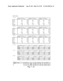 |  |
 |  |
 |  |
 |  |
 |  |
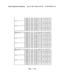 | 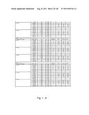 |
 |  |
 | 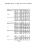 |
 |  |
 |  |
 |  |
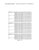 |  |
 |  |
 |  |
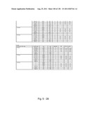 |  |
 |  |
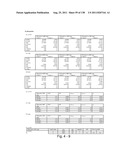 | 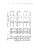 |
 |  |
 |  |
 | 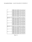 |
 | 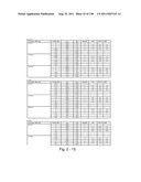 |
 | 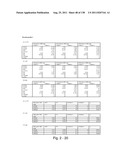 |
 | 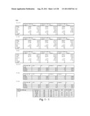 |
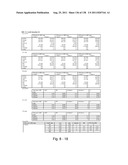 |  |
 |  |
 |  |
 |  |
 |  |
 | 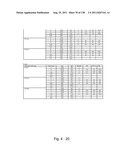 |
 |  |
 |  |
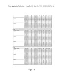 |  |
 |  |
 |  |
 | 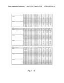 |
 |  |
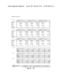 |  |
 |  |
 |  |
 |  |
 |  |
 | 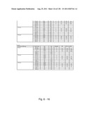 |
 |  |
 |  |
 |  |
 |  |
 |  |
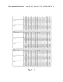 | 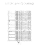 |
 |  |
 |  |
 |  |
 |  |
 |  |
 |  |
 | 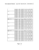 |
 | 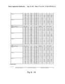 |
 | 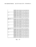 |
 |  |
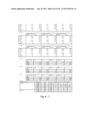 | 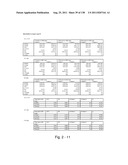 |
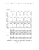 |  |
 | 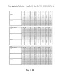 |
 |  |
 |
| New patent applications in this class: | |
| Date | Title |
|---|---|
| 2019-05-16 | Methods and uses for remotely triggered protease activity measurements |
| 2016-07-07 | Method for determining the cleaning performance of formulations |
| 2016-07-07 | Genetically encoded fret-based mmp-9 activity biosensor and use thereof |
| 2016-06-16 | Method for diagnosing cancer through detection of deglycosylation of glycoprotein |
| 2016-06-09 | Reagent for measuring endotoxin and method for measuring endotoxin |
| New patent applications from these inventors: | |
| Date | Title |
|---|---|
| 2022-09-22 | Blood biomarkers for appendicitis and diagnostics methods using biomarkers |
| 2022-07-28 | Methods and compositions for diagnosis and prognosis of renal injury and renal failure |
| 2022-07-28 | Methods for evaluation and treatment of renal injury based on c-c motif chemokine ligand 14 measurement |
| Top Inventors for class "Chemistry: molecular biology and microbiology" | |
| Rank | Inventor's name |
|---|---|
| 1 | Marshall Medoff |
| 2 | Anthony P. Burgard |
| 3 | Mark J. Burk |
| 4 | Robin E. Osterhout |
| 5 | Rangarajan Sampath |